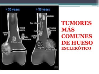
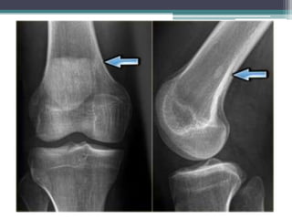
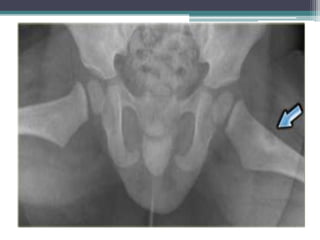
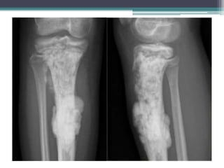
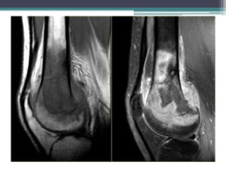
ENFERMEDAD CARACTERIZADA POR ALTERACION EN LA BIOMECANICA DE LA PATELA GENERANDO DESGASTE PROGRESIVO DE LA ARTICULACION PATELOFEMORAL , TAMBIEN CONOCIDA COMO ARTROSIS
6. ESCALA DE TOKUHASHI COLUMNA VERTEBRAL, METASTASIS EPINAL COMO VALOR PREDIC...Andres Cerquera Victoria
ESCALA DE TOKUHASHI EN COLUMNA VERTEBRAL METASTASICA COMO VALOR PREDICTIVO PARA DEFINIR MANEJO CONSERVADOR VERSUS QUIRURGICO, COMPARACION DE CLASIFICACION DE TOKUHASHI MODIFICADA T15 VERSIS CLASIFICACION ORGINAL T12
OSTEOLYSIS AND LOOSENING OF total hip arthroplasty IMPLANTS.pptx by dr vasu ...Vasu Srivastava
Aseptic Loosening of implants is caused by osteolysis. It is most significant factor limiting longevity of THA. Revision for loosening is 4x higher than next leading cause (dislocation at 13.6%), and its particularly problematic in younger patients [2].
Osteolysis is bone resorption caused by the body’s response to particulate debris generated as the THA implant wears out. Motion between any two components of the prosthesis (ie the femoral head and the acetabuluar liner, the head-neck junction of the femoral stem, or the liner and shell of the acetabulum) generates debris that floats around the joint. This debris stimulates a host response. Particles of metal, poly, or cement can all cause osteolysis, albeit different types of reaction. Osteolysis is important because it leads to implant loosening and/or periprosthetic fractures.
While osteolysis is the primary cause of loosening, infection must be part of the differential diagnosis.
Historical Perspective: Osteolysis was first described by Harris in 1976 and it was attributed to “cement disease” [3], because it was observed around the femoral component, and this was what started the drive for cementless implants. Yet after significant R&D, and development of cementless implants, osteolysis was still seen around the implants [4], and the histology was similar between cemented [5] and cementless implants [6]. Surgeons then looked for another cause of osteolysis and recognized that it was produced by wear particles.
STAGES OF OSTEOLYSIS
1) Debris production (ie poly wear) is the initial stage (we talk about metal debris in a separate section because it behaves totally differently, see section). Particulate debris in THA is produced by Abrasive and Adhesive wear (whereas the TKA produces delaminating wear: small fissures form within the poly).
▪ Adhesive wear is two surfaces bonding together causing the softer material to “peel” off as a thin film onto the harder surface during motion.
Volumetric wear is a specific type of adhesive wear, and it occurs as the femoral head articulates with the cup liner, and the amount of wear is proportional to the femoral head radius squared (therefore larger femoral head = more wear..this is why the initial Charnley implants, which used conventional poly, used a size 22 femoral head). Linear wear is caused by focused stress on a isolated part of the poly due to abnormal loading.
▪ Abrasive wear occurs when a harder surface (which is never completely smooth) cuts or ploughs through a softer surface, like a cheese grater. Both cause particle formation. Most wear occurs superiorly in the cup (or at the rim in cases of impingement).
The conventional PE wear from articulating with a Cobalt-chrome head is 0.10 mm/year. The ultramolecular weight poly (UMWPE, also known as highly-crosslinked poly) wear is about 0.02 mm/year. What is the difference between conventional and UMWPE?
Los tumores óseos constituyen uno de los diagnótico mas complicados en imagenología, lo cual nos motivo a la realización de esta presentación donde ejercitamos las principales caracteristicas radiológicas de los tumores óseos y así dar un diagnótico mas certero
la utilidad de la resonancia magnetica para la valoracion de lesiones articulares se hace imprescindible cuando evaluamos estructura blandas, ayudando a determinar un diagnostico con mayor sensibilidad. El estudio del hombro implica un conocimiento anatómico y secuencial para visualizar detalles articulares. se intenta enfocar caracteristicas imagenologicas del hombro enfocados en resonancia magnética.
Displasia Fibrosa, Encondroma y Osteoma Osteoidelgmadrid
Descripción de 3 neoplasias óseas benignas de interés en Traumatología y ortopedia y Medicina General, como parte del amplio grupo de neoplasias del tejido óseo tanto benignas como malignas.
Las malformaciones más frecuentes afectan al plastrón condroesternal.
Pectus excavatum (PEx) (tórax en embudo).
Pectus carinatum (PC) (tórax en quilla o keel chest).
Pectus arcuatum (PA) (pouter pigeon breast).
Malformaciones más infrecuentes:
Síndrome de Poland.
Síndrome de Jeune.
Distrofia torácica restrictiva adquirida.
CLASIFICACIÓN. Basada en el nivel y el número de cartílagos anormales que se insertan en el cuerpo esternal.
Tumores oseos 2. Benignos, malignos, metastasicos, diagnostico, tratamiento, manifestaciones clinicas, clasificacion, estudios de laboratorio. Quimioterapia, radioterapia, cirugia radical. Esta exposición es la original, muestra la parte 1 y 2 juntas. Esta tiene como finalidad que se muestre sola, las otras dos son para que se expongan de forma separada (proyectando), se complementan. Literatura base: Salter de TyO.
5. INFARTO ÓSEO
• TAMBIÉN CONOCIDO COMO OSTEONECROSIS ,LA
PRESENTACIÓN TÍPICA ES UNA LESIÓN CENTRAL
EN LA METÁFISIS O DIÁFISIS CON UN BORDE BIEN
DEFINIDO SERPENTIGINOSO.
• SE ASEMEJAN A LOS TUMORES CARTILAGINOSOS.
• SI LA OSTEONECROSIS SE ENCUENTRA EN LA
EPÍFISIS SE UTILIZA EL TÉRMINO DE AVASCULAR.
9. ISLA ÓSEA
• TAMBIÉN CONOCIDO COMO ENOSTOSIS, ES UNA
LESIÓN BENIGNA QUE CONSISTE EN TEJIDO ÓSEO
MADURO BIEN DIFERENCIADO DENTRO DE LA
CAVIDAD MEDULAR.
• CON FRECUENCIA ES UN HALLAZGO CASUAL,
PUDIENDO ENCONTRARSE EN CUALQUIER HUESO.
13. ENCONDROMA
• LESIÓN BENIGNA CARTILAGINOSA BASTANTE
COMÚN DE HALLAZGO CASUAL MÁS FRECUENTE
EN ADULTOS.
• PUEDE PRESENTARSE COMO :
• LESIÓN TOTALMENTE LÍTICA SIN NINGUNA
CALCIFICACIÓN.
• LESIÓN DURA CALCIFICADA.
• LESIÓN DE ASPECTO MIXTO LÍTICO CON ANILLOS,
ARCOS DE CALCIFICACIONES Y COMO
CALCIFICACIONES EN FORMA DE PALOMITAS DE
MAÍZ.
14.
15. GRANULOMA
EOSINOFÍLICO
PACIENTE DE 20 AÑOS
CON LESIÓN
ESCLERÓTICA
EXPANSIVA EN LA
CLAVÍCULA CON
PEQUEÑAS ZONAS DE
OSTEÓLISIS MAL
DEFINIDAS.
ES IMPORTANTE
DIFERENCIARLO DE LA
OSTEOMIELITIS.
16. DISPLASIA
FIBROSA
• LA DISPLASIA
FIBROSA PUEDE SER
PURAMENTE LÍTICA,
AUNQUE PUEDE
PRESENTAR
ALGUNAS
OSIFICACIONES O
CALCIFICACIONES
DE LA MATRÍZ EN
CANTIDADES
VARIABLES.
18. METÁSTASIS
• OSTEOBLÁSTICAS O METÁSTASIS ESCLERÓTICAS
DEBEN INCLUIRSE EN EL DIAGNÓSTICO
DIFERENCIAL DE CUALQUIER LESIÓN ÓSEA
ESCLERÓTICA EN PACIENTES MAYORES DE 40
AÑOS.
• ESTE TIPO DE METÁSTASIS COMUNMENTE SE
ORIGINAN EN CA. DE PRÓSTATA, MAMA Y CON
MENOR FRECUENCIA EN CA. DE PULMÓN,
LINFOMA O CARCINOIDE.
19. PACIENTE CON CA.DE
PRÓSTATA CON
NUMEROSAS
METÁSTASIS
PREDOMINANTEMEN
TE OSTEOBLÁSTICAS.
20. PACIENTE CON CA.
DE MAMA CON
METÁSTASIS
OSTEOBLÁSTICA
QUE PRESENTA
LESIÓN
ESCLERÓTICA
SUTÍL EN LA
METÁFISIS DEL
HÚMERO.
21. M.O. EN Rx. PELVIS
APENAS VISIBLE EN
HUESO ILÍACO
IZQUIERDO.
EN TC. SE OBSERVAN
LAS LESIONES CON
MEJOR DEFINICIÓN.
23. OSTEOCONDROMA
• El osteocondroma es una protuberancia ósea cubierta
por una capa cartilaginosa.
• El crecimiento de la osteocondroma tiene lugar en la
capa condroide, que corresponde con el crecimiento
normal endocondral en las placas de crecimiento. En
consecuencia, el crecimiento de los osteocondromas se
produce hasta que el paciente llega a la edad adulta.
• El crecimiento de los osteocondromas en edades adultas
(caracterizada por una gruesa capa cartilaginosa
observado como SI alta ponderado en T2 de RM); debe
plantear la sospecha de progresión a un condrosarcoma.
• El osteocondroma suele presentarse como un hallazgo
incidental, o en la forma de un síndrome con múltiples
osteocondromas.
24.
25.
26.
27. OSTEOMA OSTEOIDE
• Osteoma osteoide es una lesión ósea benigna con
un nido pequeño rodeado por una zona de
esclerosis reactiva.
• TC es muy útil para detectar y diferenciar el nido
del osteoma osteoide de otras lesiones
escleróticas como osteoblastoma, osteomielitis,
artritis, fractura de estrés y enostosis.
28. • Osteoma osteoide es una pequeña lesión (hasta 1,5
cm) osteolítica, con o sin calcificación central.
• La ubicación es generalmente cortical / excéntrico,
subperióstico a veces o incluso, intra - articular.
• Los sitios más comunes de origen incluyen el
fémur, la tibia, la espina dorsal (posterior), las
manos y los pies.
• La lesión se caracteriza por una extensa esclerosis
reactiva con engrosamiento cortical.
• TC es óptima para apreciar el nido.
• RMI también puede detectar el nido, combinado
con abundante edema de médula ósea y / o del
tejido blando. En los huesos largos, el diagnóstico
diferencial puede incluir procesos reactivos, como
fractura por estrés o absceso cortical.
29. • En la mayoría de los casos de osteoma osteoide,
el aspecto radiográfico se determina por la
esclerosis reactiva.
• En algunos casos, sin embargo el nido osteolítico
puede ser visible en la radiografía.
30.
31.
32.
33.
34. OSTEOMA
• El Osteoma consiste en tejido óseo densamente
compacto. Estas lesiones ocurren
predominantemente en la tabla externa del
neurocráneo o en un seno paranasal.
• A veces, un osteoma está presente a lo largo de la
superficie de un hueso plano o tubular. Osteoma
puede simular una displasia fibrosa fuertemente
osificada.
35.
36.
37. OSTEOMIELITIS
• Centralmente hay un área osteolítica mal
definida.
• La mayor parte de la lesión consiste en esclerosis
reactiva.
38.
39.
40.
41. OSTEOSARCOMA
Hechos y cifras
• Tumor óseo maligno primario mas común.
• Segundo tumor óseo primario más común y es
altamente maligno. Es más común entre las personas
de 10 a 25 años, aunque puede ocurrir a cualquier
edad.
• 50% alrededor de la rodilla (fémur distal con más
frecuencia que la tibia proximal) o en otros huesos
largos (como el húmero proximal), en particular en
la zona metafiso diafisaria, y puede metastatizar, por
lo general a los pulmones o en otros huesos.
• Presentación: dolor, masa, fractura patológica.
• La esclerosis está presente, ya sea en la formación de
tumores de hueso nuevo o esclerosis reactiva.
42. • Las radiografías simples típicamente revelan
lesiones con patrón apolillado o permeativo de la
zona de transición con destrucción cortical
irregular y una reacción perióstica interrumpida
con extensión de tejidos blandos.
• Una reacción perióstica conocida como “triángulo
de Codman” aparece cuando el tumor eleva el
periostio del hueso subyacente.
• La extensión cortical de tejido blando puede
producir espículas radiadas de hueso llamado
apariencia en “rayo de sol”.
43.
44.
45. • El osteosarcoma maligno produce osteoide (hueso
inmaduro) de las células óseas tumorales.
• La formación de hueso explica las imágenes
radiográficas.
• Cuando la matriz tumoral ósea es deficiente, el
osteosarcoma puede ser del subtipo telangiectasico
(que algunas veces puede imitar un quiste óseo
aneurismático), o el diagnóstico diferencial incluye
el sarcoma de Ewing o linfoma, especialmente
cuando una gran masa de tejido blando está
presente.
• En pacientes de edad avanzada, el diagnóstico
diferencial también puede ser condrosarcoma.
46. • Generalmente se desarrolla alrededor de la rodilla
• El dolor y la hinchazón son los síntomas habituales.
• Los hallazgos en los estudios de imagen varían y
pueden incluir características escleróticas y / o
líticas.
• El diagnóstico requiere una biopsia.
• Se necesitan radiografías de tórax y TC para
detectar metástasis pulmonares y una gammagrafía
ósea para detectar metástasis óseas.
47. • La RM óptima para la estadificación locorregional.
• Un protocolo de obtención de imágenes debe
incluir por lo menos una serie T1 para la
evaluación de la extensión intraósea y relación con
las articulaciones, y axial T2 fat sat, para la
evaluación de la extensión de los tejidos blandos y
la relación con importantes estructuras
neurovasculares.
• Una imagen longitudinal del hueso largo completo
se necesita para descartar metástasis no continuas
(saltarinas).
48. • El papel de las imágenes durante y después de la
quimioterapia neo adyuvante sirve para evaluar la
tasa de respuesta al tratamiento.
• En particular, la reducción significativa de la
vascularización tumoral es un fuerte indicador de
buena respuesta. Aumento de volumen del tumor
sugiere fuertemente respuesta pobre, sin embargo,
la disminución de volumen no indica
necesariamente una respuesta favorable.
49. • El osteosarcoma intraóseo de bajo grado es una
variante muy rara de osteosarcoma, que sin
embargo puede tener un aspecto radiológico
benigno y por lo tanto puede ser confundido con
otras lesiones, por ejemplo, la displasia fibrosa.
• Osteosarcomas superficiales tienen su origen en
la superficie del hueso y crecen principalmente en
los tejidos blandos circundantes, pero también
puede infiltrarse en la médula ósea. Entre estos, el
osteosarcoma parostal (o yuxtacorticales) es la
variante más frecuente, más comúnmente en el
lado posterior de la unión metafiso diafisaria distal
del fémur.
50.
51.
52.
53.
54.
55.
56.
57.
58. FRACTURAS POR ESTRÉS
• Las fracturas por estrés se producen en huesos
normales (fracturas por fatiga), o
metabólicamente debilitados (fracturas por
insuficiencia).
• Por lo general, las fracturas por estrés son fáciles
de reconocer. Con poca frecuencia puede ser
difícil de diferenciar una fractura por estrés de
una fractura patológica, que se produce en el
sitio de un tumor óseo.