SlideShare una empresa de Scribd logo
1 de 12
Descargar para leer sin conexión
Odontol Pediátr (Madrid)
Vol. 22, N.º 3, pp. 183-194, 2014
1113-5181/14/22.3/183-194
Odontología Pediátrica
Copyright © 2014 SEOP y Arán ediciones, S. L.
Caso clínico
Protocolo de actuación y tratamiento adecuado de las
complicaciones para el éxito del reimplante tras una avulsión
dental. Caso clínico
C. Gómez Ponce1
, A. Iglesias Linares2
, M.D. Huertas1
, A. Mendoza Mendoza1
1
Departamento de Estomatología. Odontopediatría. Facultad de Odontología. Universidad de Sevilla. Sevilla.
2
Departamento de Estomatología IV. Facultad de Odontología. Universidad Complutense de Madrid. Madrid
Recibido: 26-03-2014
Aceptado: 22-01-2015
Introducción
La avulsión dental supone una lesión grave en la cual
el diente se desplaza por completo fuera del alveolo
dañando los tejidos de unión y el soporte vasculo-ner-
vioso (1). La prevalencia de esta lesión varía del 0,5 %
al 16 % de todos los casos de trauma dental en dentición
permanente (2) y los dientes más frecuentemente afec-
tados son los incisivos centrales superiores (3,4) entre
los 7 y los 9 años (5). En estas edades, los dientes están
erupcionando y el desarrollo de las raíces está comple-
tándose, por lo que el ligamento está poco estructurado
RESUMEN
La prevalencia de avulsión dental supone del 0,5-16 % de
todas las lesiones traumáticas. Se presenta el tratamiento efec-
tuado a una niña de 8 años que sufrió avulsión de un incisivo
superior permanente inmaduro. El diente fue reimplantado en la
consulta odontológica después de 40 minutos en seco y 1 hora
de almacenamiento en agua. En la monitorización, a la primera
semana se detectó reabsorción radicular externa que fue tratada
con hidróxido cálcico. A los 6 meses se realizó el cierre apical
con MTAy finalmente la endodoncia de la pieza. Se describe el
caso tras 4 años de seguimiento mostrándose el diente funcional
y estético en la cavidad oral, habiéndose iniciado un tratamiento
ortodóncico a los 2 años del trauma sin observar reactivación
de la lesión. En el presente trabajo, se ilustra y enfatiza cómo
la importancia de un tratamiento inmediato y el seguimiento
de pautas clínicas protocolizadas basadas en la evidencia son
claves en el éxito del tratamiento de los dientes avulsionados.
Palabras clave: Avulsión dental. Exarticulación. Trauma
dental. Reimplantación dental.
SUMMARY
The prevalence of dental avulsion represents 0.5-16 % of
all traumatic injuries. The present study describes the treat-
ment of an 8 year-old girl who suffered an avulsion of an
immature permanent maxillary incisor. Replantation was
performed in the dental office after 40 minutes dry time and
1 hour water storage. During the first week of monitoring,
external root resorption was detected which was treated with
calcium hydroxide. At 6 months apical closure was carried out
with MTA and root canal therapy was finally performed. The
case is described with 4-year follow up of the tooth which met
the functional and aesthetic requirements in the individual.
Orthodontic treatment was started 2 years after the traumatic
injury and there was no sign of any aggravation of the injury.
This study illustrates and emphasizes the importance of imme-
diate treatment and of following an evidence-based protocol
and clinical guidelines that are key for treating avulsed teeth.
Key words: Tooth avulsion. Exarticulation. Dental trauma.
Tooth replantation.
y es flexible, además el hueso alveolar es más “elástico”,
de esta forma, los niños son más propensos a sufrir una
avulsión dental (6).
Las causas directas o factores etiológicos de este tipo
de trauma son las caídas, actividades deportivas, acciden-
tes de tráfico y la violencia (4). Además, existen facto-
res predisponentes que aumentan el riesgo de sufrir una
avulsión dental, como son la protrusión de los incisivos
superiores, un labio superior corto y antecedentes de
trauma (7).
La tasa de éxito de los dientes reimplantados oscila del
9 % al 50 % (8). Esta tasa asciende cuando los dientes
son reimplantados en condiciones favorables entre un
71 % y un 82 % (9) llegando al 95,6 % según Chappius
(10).
184	 C. Gómez Ponce ET AL.	 Odontol Pediátr
CASO CLÍNICO
Descripción y diagnóstico
Acude a la consulta de atención odontopediátrica
especializada, una niña de 8 años de edad sin patología
sistémica tras una caída en el colegio, a consecuencia de
la cual sufrió la avulsión de un incisivo central superior
derecho (Fig. 1A). Clínicamente se observaba formación
de coágulo sanguíneo (Fig. 1B) y radiográficamente el
alveolo vacío (Fig. 1C). El diente se mantuvo en una
servilleta durante 40 minutos con inclusión posterior en
agua durante una hora. Al llegar a la clínica el diente se
mostraba íntegro observándose la no finalización en el
desarrollo apical.
Protocolo de tratamiento
El diente fue reimplantado siguiendo las directrices
actuales de la IADT (11). Se anestesió a la paciente
(Mepivalem®
3 % SV, mepivacaína sin vasoconstrictor,
Dentsply), se examinó el alveolo para comprobar que
no existía obstáculo para el reimplante, se efectuó una
radiografía oclusal y otra periapical con técnica de la
bisectriz (aparato de rayos Sidexis), y se irrigó el alveolo
con suero para eliminar el coágulo sanguíneo (Fig. 2). En
este caso no se encontraron fracturas óseas pero se suturó
una laceración en tejido blando a nivel del frenillo labial.
Por otro lado, el diente se introdujo durante 5 minutos
en una solución con 20 ml de suero fisiológico + 1 mg
doxiciclina (Fig. 3) para favorecer la revascularización
y la cicatrización periodontal (12-15).
Posteriormente reimplantamos el diente con presión
digital suave y aplicamos una férula flexible o semirrí-
gida (Fig. 4A). Efectuamos una radiografía de control
para verificar la posición correcta del diente (Fig. 4B).
La férula (alambre 0,015 pulgadas tri-flex®
) se mantiene
siete días (11,12,16,17).
Se corroboró la existencia de protección inmunológica
antitetánica, asimismo se prescribió cobertura antibiótica
(50 mg/kg/día amoxicilina 7 días) (11,18). Siguiendo el
protocolo internacional se sugiere como primera opción
Fig. 2. Tras la anestesia se comprueba que no existe obstáculo
para el reimplante y se irriga el alveolo con solución salina
para eliminar el coágulo.
Fig. 1. Aspecto clínico y radiológico inicial. A. Vista frontal
del aspecto clínico inicial donde observamos ausencia del 11.
B. Vista palatina del aspecto clínico inicial donde observamos
el alveolo sangrante y vacío. C. Radiografía periapical de la
zona para verificar la ausencia de fragmentos dentales en el
alveolo o cuerpos extraños.
Fig. 3. Introducción del diente en 20 ml de suero + 1 mg
doxiciclina durante 5 minutos.
A
B
C
Vol. 22, N.º 3, 2014	 Protocolo de actuación y tratamiento adecuado de las complicaciones para el éxito	 185
	 del reimplante tras unaavulsión dental. Caso clínico
la tetraciclina (doxiciclina 2 veces al día durante una
semana con dosis adaptada para edad y peso) y como
alternativa en pacientes jóvenes con riesgo de decolora-
ción la amoxicilina o fenoximetilpenicilina (11,16).
Por último, se dieron pautas de dieta blanda durante
dos semanas e higiene con cepillo suave y enjuagues con
clorhexidina (0,1 %) 2 veces al día durante una semana
(11,12,16,19).
Monitorización clínica y radiológica
El trauma se sometió a un control clínico y radiográ-
fico periódico a la semana, 4 semanas, 3 y 6 meses, al
año, y luego anualmente durante cuatro años. La IADT
recomienda un seguimiento anual durante cinco años
(11,12,16).
En el examen radiográfico realizado a la semana se
observó una lesión por reabsorción radicular externa en
el tercio medio de la raíz (Fig. 5A). Se extirpó la pul-
pa, desbridando con limas K (Fig. 5B) e irrigando con
hipoclorito al 5 % y se introdujo hidróxido cálcico puro
hasta observar detención en la reabsorción radicular a
los 6 meses (Fig. 5C). Posteriormente se facilitó el cie-
rre apical mediante MTA (ProRoot®
MTA, Dentsply) y
se efectuó la endodoncia de la pieza con gutapercha y
sellador (AH26, Dentsply) (Fig. 5D). El caso cuenta con
un seguimiento anual durante 4 años (Figs. 6-8) obser-
vando que el diente mantiene estabilidad, funcionalidad
y estética aceptable durante el tiempo de monitorización.
DISCUSIÓN
El protocolo correcto ante una avulsión dental incluye
unos primeros auxilios en el lugar del accidente: mante-
ner la calma, manipular el diente a nivel de la corona y
Fig. 4. Reimplante y ferulización. A. Vista clínica de la
ferulización tras en reimplante. B. Verificación radiológica de
la posición correcta tras el reimplante y la ferulización.
Fig. 5. Seguimiento radiológico. A. En el seguimiento a la
semana se detectó radiológicamente lesión por reabsorción
radicular externa en el tercio medio de la raíz. B. Tratamiento
intraconducto con hidróxido de calcio para frenar la
reabsorción radicular externa. C. Detención en la reabsorción
radicular a los 6 meses. D. Cierre apical mediante MTA y
endodoncia.
A
B A
C
B
D
186	 C. Gómez Ponce ET AL.	 Odontol Pediátr
asegurarnos de que se trata de un diente permanente ya
que no es recomendable reimplantar dientes temporales
(11,16). Si el diente estuviese sucio debemos lavarlo con
agua o suero 10 segundos (11,16,21,22).
Posteriormente el tratamiento de elección es su reim-
plante (6,11,12,16,23). El reimplante inmediato (antes
de 5 minutos) asegura la viabilidad del ligamento (6,
29-27)y una curación completa (23). Todos los esfuerzos
deben hacerse para reimplantar el diente dentro de los
primeros 15-20 minutos donde es posible la cicatrización
periodontal (9,28-30). Si no fuese posible el diente debe
almacenarse en un medio adecuado (11,12,16,23). El
medio idóneo es la solución salina equilibrada de Hank
(11,12,31-33). No obstante, la disponibilidad de este tipo
de medio en el público general es irreal, con lo cual la
leche desnatada, el suero o en último término la saliva
pueden ser medios sustitutivos bastante útiles (1,11,16).
El agua, utilizada en este caso, se debe evitar (11,16,34).
Nuestro tratamiento y el pronóstico del diente es con-
dicionado por 2 factores clínicos, el desarrollo apical y la
condición de las células del ligamento (1,10-12,16,23).
Esta última depende a su vez del tiempo en seco y del
medio de almacenamiento (6,11,12,16,35,36).
Fig. 6. Aspecto clínico y radiológico al año.
Fig. 7. Aspecto clínico y radiológico a los 2 años.
A
A
B
B
Respecto al desarrollo apical es importante cono-
cer que sólo en dientes inmaduros es posible esperar la
revascularización pulpar, por lo que sólo en estos casos
no efectuaremos el tratamiento de conductos –salvo que
existan indicios de necrosis pulpar (11,16) o se exce-
dan los 60 minutos en seco (1,11,16). Para favorecer la
revascularización se introduce el diente en doxiciclina
o minociclina 0,05 % (1 mg/20 ml) 5 minutos antes
del reimplante (11-16). En dientes con desarrollo apical
maduro siempre efectuaremos tratamiento de conductos.
El momento del tratamiento de conductos depende del
periodo extraoral (11,12,16,37) y el hidróxido de calcio
es el medicamento de elección (11).
La viabilidad del ligamento es el otro factor a con-
siderar. En este caso, estaba disminuida puesto que a
los 30 minutos en seco se produce la muerte del 50 %
de las células y a la hora apenas quedan células viables
(38). Para aumentar la cicatrización periodontal debemos
introducir el diente antes del reimplante en 1 mg minoci-
clina o doxiciclina/20 ml solución salina 5 minutos. Sin
embargo, cuando el tiempo extraoral en seco es superior
a 60 minutos las células están necróticas y son invia-
bles (23,28,30), debemos entonces eliminar el ligamento
(11,12,16,23,39) y esperar la reabsorción sustitutiva y
Vol. 22, N.º 3, 2014	 Protocolo de actuación y tratamiento adecuado de las complicaciones para el éxito	 187
	 del reimplante tras unaavulsión dental. Caso clínico
anquilosis (11,16). El objetivo del reimplante en estos
casos es estético, funcional, por razones psicológicas y
para mantener el hueso alveolar (11,16,39). Para reducir
la velocidad de reemplazo óseopueden introducirse estos
dientes antes de su reimplantación en una solución al
2 % de fluoruro de sodio 20 minutos (11).
La condición de las células del ligamento también
pudo verse afectada por el medio de almacenamiento.
El agua tiene baja osmolaridad (28,40), al ser hipotóni-
ca provoca la lisis celular (41) y el aumento de la infla-
mación en el reimplante (34). Según Andreasen (6) el
almacenamiento en agua más de 20 minutos siempre dará
como resultado reabsorción radicular, pero este mismo
autor mostró cómo dientes reimplantados después de
largos periodos extraorales no mostraron reabsorción
radicular (44); también mostró un caso en el que dos
incisivos que habían estado fuera de la boca 16 horas
permanecieron funcionales 2 años. Shiu-yin Cho (45)
mostró funcionalidad y estética durante 2 años de un
diente que estuvo en seco 18 horas. Khalilak (46)descri-
bió estabilidad durante 5 años de un diente reimplantado
tras 270 minutos. Siguiendo estas recomendaciones el
diente fue reimplantado después de una hora y cuarenta
minutos extraorales considerando comprometida la via-
bilidad del ligamento pero corroborando tras 4 años de
seguimiento que el diente permanece funcional y estético
en la cavidad oral.
Adicionalmente, el caso descrito aborda una pieza
dental inmadura donde había que sopesar el intento de
revascularización frente a la posible aparición de com-
plicaciones debido al largo periodo extraoral.
Dos factores son críticos para que la revascularización
pueda tener éxito, el tiempo (26,47) y la presencia de
bacterias (26,47,48), ambos factores comprometidos en
nuestro caso. Respecto al tiempo Kling (47)y Andreasen
(26) consideran posible la revascularización si el dien-
te se reimplanta dentro de los 45 minutos posteriores
al trauma. Cvek (48) y otros autores (26,47) describen
que la ocurrencia de revascularización es altamente
dependiente de la presencia o ausencia de bacterias en
la pulpa (13,15). En la actuación de dicho caso se intentó
favorecer la revascularización introduciendo el diente en
20 ml de solución salina y 1 mg de doxiciclina (11). No
consiguió la revascularización en este caso, pero pudo
contribuir a la atenuación de la reabsorción radicular y
a la curación periodontal. Además, el tratamiento sisté-
mico antibiótico también ayudó a evitar la proliferación
bacteriana y limitar la reabsorción inflamatoria (49,50).
En el control radiológico a la semana se detectó reab-
sorción radicular inflamatoria externa en el tercio medio de
la raíz. Este tipo de reabsorción está asociada con necrosis
e infección de la pulpa, de forma que las bacterias entran
en el espacio periodontal a través de los túbulos dentina-
rios (4) y junto al daño en el cemento resulta en reabsor-
ción inflamatoria externa (51). Actualmente el tratamiento,
prevención o limitación de la reabsorción radicular incluye
la terapia con hidróxido de calcio (52), aunque también
existen buenos resultados con una combinación de antibió-
tico y corticoide como Ledermix (53-56). Estudios actua-
les plantean el posible uso de corticoides (57), calcitonina
(58), acetazolamida (59), bifosfonatos (60,61)o derivado
de la matriz del esmalte (62,63). Sin embargo, hoy día, el
hidróxido cálcico sigue siendo el medicamento de elección
y fue el que usamos en el presente caso. Posteriormente
se facilitó el cierre apical mediante MTA y finalmente se
efectuó la endodoncia de la pieza.
CONCLUSIONES
Reimplantar el diente a la mayor brevedad posible o
mantenerlo en un medio adecuado son parámetros clí-
nicos fundamentales en el pronóstico de la pieza avul-
sionada.
Fig. 8. A. Aspecto radiológico a los 3 años y medio durante el movimiento ortodóncico. B. Seguimiento radiológico a los 4 años. El
diente se mantuvo estable funcional y estéticamente aceptable. No se observó reabsorción inflamatoria ni anquilosis.
A B
188	 C. Gómez Ponce ET AL.	 Odontol Pediátr
El estado del ápice y el periodo extraoral en seco
condicionan dramáticamente el pronóstico del diente
avulsionado.
El seguimiento de pautas clínicas protocolizadas basa-
das en la evidencia determinan en gran medida el éxito
del tratamiento a pesar de su mal pronóstico inicial.
BIBLIOGRAFÍA
1.	 Trope M. Clinical management of the avulsed tooth: present
strategies and future directions. Dent Traumatol 2002;18:1-11.
2.	 Davidovich E, Moskovitz M, Moshonov J. Replantation of an
immature permanent central incisor following preeruptive trau-
matic avulsion. Dent Traumatol 2008;24(5):e47-52.
3.	 Gonda F, Nagase M, Chem RB, Yakata H, Nakajima T. Replan-
tation: An analysis of 29 teeth. Oral Surg Oral Med Oral Pathol
1990;70:650-5.
4.	 Tzigkounakis V, Merglová V, Hecová H, Netolický J. Retrospec-
tive clinical study of 90 avulsed permanent teeth in 58 children.
Dent Traumatol 2008;24:598-602.
5.	 Baginska J, Wilczynska-Borawska M. First-aid algorithms in
dental avulsion. J Sch Nurs 2012;28(2):90-4.
6.	 Andreasen JO, Borum, MK, Jacobsen HL, Andreasen FM.
Replantation of 400 avulsed permanent incisors. 4. Factors relat-
ed to periodontal ligament healing. Endodontics Dent Traumatol
1995;11:76-89.
7.	 Brin I, Ben-Bassat Y, Heling I, Brezniak N. Profile of an ortho-
dontic patient at risk of dental trauma. Endod Dent Traumatol
2000;16:111-5.
8.	 Steiner DR. Avulsed maxillary central incisors: The case for
replantation. Am J Orthod Dentofacial Orthop 2012;142(1):8,
10, 12, 14, 16.
9.	 Andersson L, Bodin I. Avulsed human teeth replanted within
15 minutes —a long-term clinical follow-up study. Endod Dent
Traumatol 1990;6:37-42.
10.	 Chappuis V, von Arx T. Replantation of 45 avulsed permanent
teeth: A 1-year follow-up study. Dent Traumatol 2005;21(5):
289-96.
11.	 Andersson L, Andreasen JO, Day P, Heithersay G, Trope M,
Diangelis AJ, et al. International Association of Dental Trau-
matology guidelines for the management of traumatic den-
tal injuries: 2. Avulsion of permanent teeth. Dent Traumatol
2012;28(2):88-96.
12.	 Flores MT, Andreasen JO, Bakland LK, Feiglin B, Gutmann JL,
Oikarinen K et al.; International Association of Dental Trauma-
tology. Guidelines for the evaluation and management of trau-
matic dental injuries. Dent Traumatol 2001;17:193-8.
13.	 Cvek M, Cleaton-Jones P, Austin J, Lownie J, Kling M, Fatti P.
Effect of topical application of doxycycline on pulp revascular-
ization and periodontal healing in reimplanted monkey incisors.
Endod Dent Traumatol 1990;6(4):170-6.
14.	 Yanpiset K, Trope M. Pulp revascularization of replanted imma-
ture dog teeth after different treatment methods. Endod Dent
Traumatol 2000;16:211-7.
15.	 Ritter AL, Ritter AV, Murrah V, Sigurdsson A, Trope M. Pulp
revascularization of replanted immature dog teeth after treat-
ment with minocycline and doxycycline assessed by laser Dop-
pler flowmetry, radiography, and histology. Dent Traumatol
2004;20:75-84.
16.	 Flores MT,Andersson L,Andreasen JO, Bakland LK, Malmgren
B, Barnett F. Guidelines for the management of traumatic den-
tal injuries. II. Avulsion of permanent teeth. Dent Traumatol
2007;23:130-6.
17.	 Andreasen JO, Friskopp J, Blomloff L. Fiberglass splinting of
traumatized teeth. J Dent Child 1983;3:21-24.
18.	 Hammarstrom L, Blomloff L, Feiglin B, Andersson L, Lindskog
S. Replantation of teeth and antibiotic treatment. Endod Dent
Traumatol 1986;2:51-7.
19.	 Ram D, Cohenca N. Therapeutic protocols for avulsed permanent
teeth: Review and clinical update. Pediatr Dent 2004;26:251-5.
20.	 Davidovich E, Moskovitz M, Moshonov J. Replantation of an
immature permanent central incisor following pre-eruptive trau-
matic avulsion. Dent Traumatol 2008;24:e47-e52.
21.	 Nesiama JO, Sinn DP. Tooth avulsion. Clinical Pediatric Emer-
gency Medicine 2010;11:108-11.
22.	 Emerich K, Kaczmarek J. First aid for dental trauma caused by
sports activities. State of knowledge, treatment and prevention.
Sports Med 2010;40(5):361-6.
23.	 Trope M. Avulsion of permanent teeth: Theory to practice. Den-
tal Traumatology 2011;27(4):281-94.
24.	 Andreasen JO, Borum MK, Jacobsen HL, Andreasen FM.
Replantation of 400 avulsed permanent incisors. 1. Diagnosis
of healing complications. Endod Dent Traumatol 1995;11:51-8.
25.	 Andreasen JO, Borum MK, Jacobsen HL, Andreasen FM.
Replantation of 400 avulsed permanent incisors. 2. Factors
related to pulpal healing. Endod Dent Traumatol 1995;11:59-68.
26.	 Andreasen JO, Borum MK, Jacobsen HL, Andreasen FM.
Replantation of 400 avulsed permanent incisors. 3. Factors
related to root growth. Endod Dent Traumatol 1995;11:69-75.
27.	 Kinirons MJ, Gregg TA, Welbury RR, Cole BOI. Variations in
the presenting and treatment features in reimplanted permanent
incisors in children and their effect on the prevalence of root
resorption. Br Dent J 2000;189:263-6.
28.	 Andreasen JO. The effect of extra-alveolar period and stor-
age media upon periodontal and pulpal healing after replanta-
tion of mature permanent incisors in monkeys.Int J Oral Surg
1981;10:43-53.
29.	 Barrett EJ, Kenny DJ. Avulsed permanent teeth: A review of
the literature and treatment guidelines. Endod Dent Traumatol
1997;13:153-63.
30.	 Soder PO, Otteskog P, Andreasen JO, Modeer T. Effect of dry-
ing on viability of periodontal membrane. Scand J Dent Res
1977;85:167-72.
31.	 Hiltz J, Trope M. Vitality of human lip fibroblasts in milk. Hank’s
balanced salt solution and Via Span storage media. Endod Dent
Traumatol 1991;7:69-72.
32.	 Ashkenazi M, Sarnat H, Keila S. In vitro viability, mitogenic-
ity and clonogenic capacity of periodontal ligament cells after
storage in six different media. Endod Dent Traumatol 1999;15:
149-56.
33.	 Trope M, Friedman S. Periodontal healing of replanted dog teeth
stored in Viaspan, milk and Hanks Balanced Salt Solution. Endod
Dent Traumatol 1992; 8:183-8.
34.	 Blomlof L. Milk and saliva as possible storage media for trau-
matically exarticulated teeth prior to replantation. Swed Dent J
Suppl 1981;8:1-26.
35.	 Pettiette M, Hupp J, Mesaros S, Trope M. Periodontal healing of
extracted dogs’teeth air-dried for extended periods and soaked in
various media. Endod Dent Traumatol 1997;13:113-8.
36.	 Lin DG, Kenny DJ, Barrett EJ, Lekic P, McCulloch CAG. Stor-
age conditions of avulsed teeth affect the phenotype of cultured
human periodontal ligament cells. J Periodont Res 2000;35:
42-50.
37.	 Giannetti L, Murri A. Clinical evidence and literature to com-
pare two different therapeutic protocols in tooth avulsion. Eur J
Pediatr Dent 2006;7:122-30.
38.	 Flogia I. Avulsión dentaria. Otros recursos terapéuticos. EJER
2012;11(1):575-96.
39.	 Panzarini SR, Gulinelli JL, Poi WR, Sonoda CK, D Pedrini,
Brandini DA. Treatment of root surface in delayed tooth replan-
tation:Areview of literature. Dent Traumatol 2008;24(3):277-82.
40.	 Sigalas E, Regan JD, Kramer PR, Witherspoon DE, Opperman
LA. Survival of human periodontal ligament cells in media pro-
posed for transport of avulsed teeth. Dent Traumatol 2004;20:21-8.
41.	 Marino TG, West LA, Liewehr FR, Mailhot JM, Buxton TB,
Runner RR, et al. Determination of periodontal ligament cell
viability in long shelf-life milk. J Endod 2000;26:699-702.
Correspondencia:
Cristina Gómez Ponce
Departamento de Estomatología
Odontopediatría
Facultad de Odontología
Universidad de Sevilla
e-mail: cristinagomezponce@hotmail.com
Vol. 22, N.º 3, 2014	 Protocol of action and appropriate treatment of complications for replantation 	 189
	 success following dental avulsion. A case report
Protocol of action and appropriate treatment of complications
for replantation success following dental avulsion. A case report
C. Gómez Ponce1
, A. Iglesias Linares2
, M.D. Huertas1
, A. Mendoza Mendoza1
1
Department of Dentistry. Pediatric Dentistry. Faculty of Dentistry. University of Seville. Seville, Spain. 2
Department
of Dentistry IV. Faculty of Dentistry. Universidad Complutense de Madrid. Madrid, Spain
Clinical case
42.	 Pohl Y, Tekin U, Boll M, FilippiA, Kirschner H. Investigation on
a cell culture medium for storage and transportation of avulsed
teeth. Aust Endod J 1999;25:70-5.
43.	 Andersson L, Bodin I, Sorensen S. Progression of root resorption
following replantation of human teeth after extended extraoral
storage. Endod Dent Traumatol 1989;5:38-47.
44.	 Andreasen JO, Hjorting-Hansen E. Replantation of teeth. I.
Radiographic and clinical study of 110 human teeth replanted
after accidental loss. Acta Odontol Stand 1966;24:263-86.
45.	 Cho SY, Cheng AC. Replantation of an avulsed incisor after
prolonged dry storage: A case report. J Can Dent Assoc
2002;68(5):297-300.
46.	 Khalilak Z, Shikholislami M, Mohajeri L. Delayed tooth replan-
tation after traumatic avulsion: A case report. Iran Endod J
2008;3(3):86-9.
47.	 Kling M, Cvek M, Mejare I. Rate and predictability of pulp
revascularization in therapeutically reimplanted permanent inci-
sors. Endod Dent Traumatol 1986;2(3):83-9.
48.	 Cvek M, Cleaton-Jones P, Austin J, Lownie J, Kling M, Fatti P.
Pulp revascularization in reimplanted immature monkey incisors
– predictability and the effect of antibiotic systemic prophylaxis.
Endod Dent Traumatol 1990;6:157-69.
49.	 Hammarstrom L, Pierce A, Blomlof L, Feiglin B, Lindskog S.
Tooth avulsion and replantation: A review. Endod Dent Trau-
matol 1986;2:1-9.
50.	 Sae-Lim V, Wang CY, Trope M. Effect of systemic tetracicline
and amoxicillin on inflammatory root resorption of replanted
dog, teeth. Endod Dent Traumatol 1998;14:216-20.
51.	 Tronstad L. Root resorption etiology, terminology and clinical
manifestations.Endod Dent Traumatol 1988;4:241.
52.	 Barnett F. The role of endodontics in the treatment of luxated
permanent teeth. Dent Traumatol 2002;18(2):47-56.
53.	 Bryson EC, Levin L, Banchs F, Abbott PV, Trope M. Effect of
immediate intracanal placement of Ledermix Paste(R) on healing
of replanted dog teeth after extended dry times. Dent Traumatol
2002;18:316-21.
54.	 Wong KS, Sae-Lim V. The effect of intracanalLedermix on root
resorption of delayed-replanted monkey teeth. Dent Traumatol
2002;18:309-15.
55.	 Pierce A, Lindskog S. The effect of an antibiotic corticoste-
roid combination on inflammatory root resorption. J Endod
1988;14:459-64.
56.	 Thong YL, Messer HH, Siar CH, Saw LH. Periodontal response
to two intracanal medicaments in replanted monkey incisors.
Dent Traumatol 2001;17:2549.
57.	 Kirakozova A, Teixeira FB, Curran AE, Gu F, Tawil PZ, Trope
M. Effect of intracanal corticosteroids on healing of replanted
dog teeth after extended dry times. J Endod 2009;35:663-7.
58.	 Pierce A, Berg JO, Lindskog S. Calcitonin as an alternative ther-
apy in the treatment of root resorption. J Endod 1988;14:459-65.
59.	 Mori GG, Garcia RB. Microscopic study of the effect of rootsur-
face treatment with acetazolamida in avulsed and reimplanted rat
teeth. Rev Fac Odontol Bauru 2002;10:180-5.
60.	 Lustrosa-Pereira A, Garcia RB, de Moraes IG, Bernadineli N,
Bramante CM, Bortoluzzi EA. Evaluation of the topical effect
of alendronate on the root surface of extracted and replant-
ed teeth. Microscopic analysis on rats’ teeth. Dent Traumatol
2006;22:30-5.
61.	 Levin L, Bryson EC, Caplan D, Trope M. Effect of topical alen-
dronate on root resorption of dried replanted dog teeth. Dent
Traumatol 2001;17:120-6.
62.	 Schjott M, Andreasen JO. Emdogain does not prevent progres-
sive root resorption after replantation of avulsed teeth: A clinical
study. Dent Traumatol 2005;21:46-50.
63.	 Fillippi A, Pohl Y, von Arx T. Treatment of replacement resorp-
tion by intentional replantation, resection of the ankylosedsites,
and Emdogain – results of a 6-year survey. Dent Traumatol
2006;22:307-11.
RESUMEN
La prevalencia de avulsión dental supone del 0,5-16 % de
todas las lesiones traumáticas. Se presenta el tratamiento efec-
tuado a una niña de 8 años que sufrió avulsión de un incisivo
superior permanente inmaduro. El diente fue reimplantado en la
consulta odontológica después de 40 minutos en seco y 1 hora
de almacenamiento en agua. En la monitorización, a la primera
semana se detectó reabsorción radicular externa que fue tratada
con hidróxido cálcico. A los 6 meses se realizó el cierre apical
con MTAy finalmente la endodoncia de la pieza. Se describe el
caso tras 4 años de seguimiento mostrándose el diente funcional
y estético en la cavidad oral, habiéndose iniciado un tratamiento
ortodóncico a los 2 años del trauma sin observar reactivación
de la lesión. En el presente trabajo, se ilustra y enfatiza cómo
la importancia de un tratamiento inmediato y el seguimiento
de pautas clínicas protocolizadas basadas en la evidencia son
claves en el éxito del tratamiento de los dientes avulsionados.
Palabras clave: Avulsión dental. Exarticulación. Trauma
dental. Reimplantación dental.
SUMMARY
The prevalence of dental avulsion represents 0.5-16 % of
all traumatic injuries. The present study describes the treat-
ment of an 8 year-old girl who suffered an avulsion of an
immature permanent maxillary incisor. Replantation was
performed in the dental office after 40 minutes dry time and
1 hour water storage. During the first week of monitoring,
external root resorption was detected which was treated with
calcium hydroxide. At 6 months apical closure was carried out
with MTA and root canal therapy was finally performed. The
case is described with 4-year follow up of the tooth which met
the functional and aesthetic requirements in the individual.
Orthodontic treatment was started 2 years after the traumatic
injury and there was no sign of any aggravation of the injury.
This study illustrates and emphasizes the importance of imme-
diate treatment and of following an evidence-based protocol
and clinical guidelines that are key for treating avulsed teeth.
Key words: Tooth avulsion. Exarticulation. Dental trauma.
Tooth replantation.
190	 C. GÓMEZ PONCE ET AL.	 Odontol Pediátr
INTRODUCTION
Tooth avulsion is a serious injury during which a tooth
is completely displaced from the alveolus, attachment
damage occurs and the neurovascular bundle is affected
(1). The prevalence of this lesion varies between 0.5 %
and 16 % of all dental trauma cases related to the perma-
nent dentition (2) and the teeth that are more commonly
affected are the upper central incisors (3,4) between the
ages of 7 and 9 years (5).At this age the teeth have erupted
but root development is not quite complete. The periodon-
tal ligament lacks some structure and it is flexible and, in
addition, the alveolar bone is more “elastic”. Because of
this children are more inclined to suffer the avulsion of a
tooth (6). The direct causes or etiological factors behind
this type of traumatic injury are falls, sporting activities,
traffic accidents and violence (4). In addition there are pre-
disposing factors that increase the risk of suffering tooth
avulsion such as protrusion of the upper incisors, short
lower lip, and antecedents of trauma (7).
The success rate of replanted teeth ranges between
9 % and 50 % (8). This rate increases when the teeth are
replanted in favorable conditions to 71 % and 82 % (9)
even reaching 95.6 % according to Chappius (10).
CASE REPORT
Description and diagnosis
An 8 year-old girl, with no systemic disease, attended
the specialized pediatric dentistry department after a fall
at school involving the avulsion of the upper right central
incisor (Fig. 1A). The clinical examination revealed the
formation of a blood clot (Fig. 1B) and the radiographic
findings showed an empty alveolus (Fig. 1C). The tooth
had been kept in a napkin for 40 minutes and it was later
placed in water for an hour. On arrival at the clinic the
tooth was intact and incomplete apical closure could be
observed.
Treatment protocol
The tooth was reimplanted following the current
guidelines of the IADT (11). The patient was anesthe-
tized with (3 % Mepivalem®
SV, mepivacaine without
vasoconstrictor, Dentsply). The alveolus was examined
in order to verify that no obstacles would be encountered
during the replantation and an occlusal radiograph and
another periapical radiograph were carried out using the
bisecting angle technique (Sidexis X-ray machine). The
alveolus was irrigated with saline in order to eliminate
the blood clot (Fig. 2). Bone fractures were not found but
a laceration of the soft tissues by the labial frenulum was
sutured. The tooth was placed for 5 minutes in a solu-
tion of 20 ml of saline with 1 mg of doxycycline (Fig. 3)
in order to encourage revascularization and periodontal
healing (12-15).
The tooth was then reimplanted with gentle finger
pressure and a flexible or semi-rigid splint was fitted
(Fig. 4A). A control radiograph was taken in order to
verify the correct position of the tooth (Fig. 4B). The
Fig. 2. Following anesthesia, the area was checked to ensure there
was nothing hampering the replantation. The alveolar socket was
irrigated with saline solution in order to eliminate the blood clot.
Fig. 1. Initial clinical and radiologic findings. A. Frontal view
of initial clinical appearance showing missing 11; B. Palatal
view of initial clinical appearance showing bleeding alveolar
socket. C. Periapical radiograph of the area in order to ensure
no tooth fragment or foreign body in the alveolar socket.
A
B
C
Vol. 22, N.º 3, 2014	 Protocol of action and appropriate treatment of complications for replantation 	 191
	 success following dental avulsion. A case report
splint (0.015 inch tri-flex®
wire) was kept in place for a
week (11,12,16,17).
Her anti-tetanus status was checked, and antibiotic
was prescribed (50 mg/kg/day amoxicillin for a week)
(11,18). The international protocol suggests tetracycline
as a first option (doxycycline twice a day for a week with
the dosage adapted to age and weight) and for young
patients at risk of discoloration amoxicillin or phenoxy-
methylpenicillin (11,16).
Lastly, instructions were given regarding a soft diet
for two weeks and hygiene with a soft toothbrush and
chlorhexidine rinses (0.1 %) twice a day for a week
(11,12,16,19).
Clinical monitoring and radiology
The traumatic injury was monitored clinically and
radiographically over regular periods: At one week,
4 weeks, 3 and 6 months and at one year, and then annu-
ally for four years. The IADT recommends an annual
follow-up of five years (11,12,16).
During the radiographic examination a week later a
lesion was observed as a result of external root resorp-
tion in the mid-third of the root (Fig. 5A). The pulp was
extracted, debridement was performed with a K-file
(Fig. 5B) and it was irrigated with 5 % hypochlorite.
Pure calcium hydroxide was introduced until the root
resorption was arrested at 6 months (Fig. 5C).
Apical closure was then achieved with MTA (Pro-
Root®
MTA, Dentsply) and root canal treatment was car-
ried out with gutta-percha and a sealant (AH26, Dents
ply) (Fig. 5D). The case has had an annual follow-up
of 4 years (Figs. 6-8) and the tooth has shown stability
and functionality during monitoring, and an acceptable
aesthetic appearance.
DISCUSSION
The correct protocol for an avulsed tooth includes first
aid at the site of the accident: Keeping calm, handling the
tooth by the crown and ensuring that it is a permanent
tooth, as the replantation of primary teeth is not recom-
mendable (11,16). If the tooth is dirty it should be cleaned
with water or saline for 10 seconds (11,16,21,22).
After this the treatment of choice is replantation
(6,11,12,16,23). Immediate replantation (after 5 minutes)
ensures the viability of the ligament (6,29-27) and complete
healing (23). Every effort should be made to reimplant the
tooth within the first 15-20 minutes to facilitate periodontal
healing (9,28-30). If not possible the tooth should be stored
in a suitable media (11,12,16,23). The ideal medium is
Hanks balanced salt solution (11,12,31-33). Nevertheless,
the availability of this type of medium among the general
public is unrealistic, and skimmed milk, saline or as a last
resort saliva can be quite useful substitutes (1,11,16). Water
in this case should be avoided (11,16,34). The treatment
given and the prognosis of the tooth is conditioned by two
clinical factors, root development stage and the condition
of the ligament cells (1,10-12,16,23). The latter depends on
the dry time and the storage medium (6,11,12,16,35,36).
Fig. 3. The tooth was placed in 20 ml of saline solution + 1 mg
doxycycline for 5 minutes.
Fig. 4. Replantation and splinting. A. Clinical appearance
of splinting after the replantation. B. correct position after
replantation and splinting is verified radiographically.
A
B
192	 C. GÓMEZ PONCE ET AL.	 Odontol Pediátr
With regard to root development stage, it is important
to be aware that with immature teeth it is possible to
wait for pulp revascularization, and only in these cases
should root canal therapy not be carried out (unless there
is evidence of pulp necrosis (11,16) or if there is dry time
of over 60 minutes (1,11,16). In order to promote revas-
cularization, the tooth should be introduced in doxycy-
cline or minocycline 0.05 % (1 mg/20 ml) for 5 minutes
before replantation (11-16). Root canal therapy should
always be carried out in teeth with mature apexes. The
extraoral period (11,12,16,37) will determine when root
canal therapy is carried out, and calcium hydroxide is the
treatment of choice (11).
The viability of the ligament is another factor to take
into consideration. In the present case it was reduced
because after 30 minutes dry time, 50 % of the cells die
and after an hour there will hardly be any viable cells
at all (38). In order to encourage periodontal healing,
the tooth should be introduced before the replantation
into 1 mg of minocycline or doxycycline/20 ml of saline
solution for 5 minutes. However, when the extraoral dry
time is over 60 minutes the cells will be necrotic and non-
viable (23,28,30), and the ligament should be eliminated
(11,12,16,23,39) as replacement resorption and ankylosis
will ensue (11,16). The aim of the replantation in these
cases is aesthetic and functional, due to psychological
reasons and to preserve the alveolar bone (11,16,39). In
order to reduce the rate of bone replacement, these teeth
can be placed in a solution of 2 % sodium fluoride for
20 minutes (11).
The condition of the ligament cells may also have
been affected by the storage medium. Water has low
osmolarity (28,40), and the hypotonic effect will lead to
cell lysis (41) and increased inflammation of the replan-
tation (34). According to Andreasen (6) water storage of
more than 20 minutes will always lead to root resorption,
but this same author showed how replanted teeth after a
extended extraoral periods did not result in induced root
resorption (44). He also reported a case in which two
Fig. 5. Radiological follow-up. A. During follow-up at one
week, the lesion was detected radiologically due to external
root resorption in the mid-third of the root. B. Root canal
treatment with calcium hydroxide in order to arrest external
root resorption. C. Arrested root resorption at 6 months. D.
Closure of the apex using MTA and root canal therapy.
A
C
B
D
Fig. 6. Clinical and radiographic appearance at one year.
A
B
Vol. 22, N.º 3, 2014	 Protocol of action and appropriate treatment of complications for replantation 	 193
	 success following dental avulsion. A case report
Fig. 7. Clinical and radiographic appearance at two years.
incisors that had been outside the mouth for 16 hours
remained functional for two years. Shiu-yin Cho (45)
demonstrated the functionality and aesthetic appearance
for 2 years of a tooth that had been kept dry for 18 hours.
Khalilak (46) described stability over 5 years of a tooth
that was replanted after 270 minutes. Following these
recommendations the tooth was replanted after one hour
and forty minutes extraoral time although the viability
of the ligament was considered compromised but, after
a follow-up of four years the tooth showed functional
stability and it had an acceptable aesthetic appearance
in the oral cavity.
Additionally, the case described concerned an imma-
ture tooth where the attempt to revascularize had to be
weighed up against the possible appearance of complica-
tions due to the extended extraoral time.
Two factors are critical for revascularization to be
successful, time (26,47) and the presence of bacteria
(26,47,48), and both factors but our casein jeopardy.
With regard to time, Kling (47) and Andreasen (26)
consider that revascularization is possible if the tooth
is replanted within 45 minutes of the trauma. Cvek (48)
and other authors (26,47) showed that pulp revasculariza-
tion is highly dependent upon the presence or absence of
bacteria in the pulp (13,15). In this case of ours, revascu-
larization was encouraged by placing the tooth in 20ml
of saline solution and 1 mg of doxicyline (11). Revascu-
larization was not achieved in this case but it may have
contributed to the attenuation of root resorption and peri-
odontal healing. In addition systemic antibiotic treatment
also helped to avoid bacterial proliferation and to limit
inflammatory root resorption (49,50).
Monitoring by X-ray a week later revealed external
inflammatory root resorption at the mid-third of the root.
This type of resorption is associated with necrosis and pulp
infection, as bacteria enter the periodontal space through the
dentinal tubules (4) and, with the damage to the cementum,
Fig. 8. A. Radiographic appearance at 3 years and a half during orthodontic movement. B. Radiological follow-up at 4 years. The tooth
remained stable and functional and it had an aesthetically acceptable appearance. There was no inflammatory resorption or ankylosis.
A B
A
B
194	 C. GÓMEZ PONCE ET AL.	 Odontol Pediátr
there is external inflammatory resorption (51). Currently the
treatment, prevention or control of root resorption includes
calcium hydroxide therapy (52), although there are also
good results with a combination of antibiotics and corti-
costeroid such as Ledermix (53-56). Current studies raise
the possibility of using corticosteroids (57), calcitonine (58),
acetazolamide (59), bisphosphonates (60,61) or enamel
matrix derivatives (62,63). However, calcium hydroxide
continues being the medication of choice and it was used
in this case study. Closure of the root apex was carried out
with MTA and lastly root canal therapy was performed.
CONCLUSIONS
Replantation of the tooth as quickly as possible, or
maintaining it in suitable media, are basic clinical param-
eters for the prognosis of an avulsed tooth.
The condition of the apex and the extraoral dry time
have a dramatic effect on the prognosis of the avulsed
tooth.
Following evidence-based clinical guidelines that are
protocolized will determine to a large extent the success
of the treatment despite the initial prognosis being poor.

Más contenido relacionado

La actualidad más candente

Caso clínico: Caries dentinaria profunda
Caso clínico: Caries dentinaria profundaCaso clínico: Caries dentinaria profunda
Caso clínico: Caries dentinaria profundaJorge Araneda Arteaga
 
Formato caso clinico_clinica_estomatologica_pediatrica
Formato caso clinico_clinica_estomatologica_pediatricaFormato caso clinico_clinica_estomatologica_pediatrica
Formato caso clinico_clinica_estomatologica_pediatricaAntony Ayala Salazar
 
Presentación ficha miguel lagos completa Prótesis Removible
Presentación ficha miguel lagos completa Prótesis RemoviblePresentación ficha miguel lagos completa Prótesis Removible
Presentación ficha miguel lagos completa Prótesis RemovibleJuvenilBaez
 
Caso clínico Paulina Espinoza
Caso clínico Paulina EspinozaCaso clínico Paulina Espinoza
Caso clínico Paulina EspinozaPaulina Espinoza L
 
Caso Clínico Integral
Caso Clínico IntegralCaso Clínico Integral
Caso Clínico IntegralJuvenilBaez
 
Caso Clinico de Geriatria
Caso Clinico de Geriatria Caso Clinico de Geriatria
Caso Clinico de Geriatria Linette Montufar
 
Caso clinico unfv-2 - tratamiento - nestor tafur chavez - odontologia - pro...
Caso clinico   unfv-2 - tratamiento - nestor tafur chavez - odontologia - pro...Caso clinico   unfv-2 - tratamiento - nestor tafur chavez - odontologia - pro...
Caso clinico unfv-2 - tratamiento - nestor tafur chavez - odontologia - pro...Nestor Tafur Chávez
 
Cg 2189 Vega Jeseily Tpi 2009 2
Cg 2189 Vega Jeseily Tpi 2009 2Cg 2189 Vega Jeseily Tpi 2009 2
Cg 2189 Vega Jeseily Tpi 2009 2Milagros Daly
 
Sindrome sjogrens implanto, Patología Oral.
Sindrome sjogrens implanto, Patología Oral.Sindrome sjogrens implanto, Patología Oral.
Sindrome sjogrens implanto, Patología Oral.Dr Reyna
 
Caso clínico Dra Paty, by Luis & Juan
Caso clínico Dra Paty, by Luis & Juan Caso clínico Dra Paty, by Luis & Juan
Caso clínico Dra Paty, by Luis & Juan diportzaragoza
 
Plan de tratamiento integral
Plan de tratamiento integralPlan de tratamiento integral
Plan de tratamiento integralLa Turca
 
Radiología de pacientes con necesidades especiales
Radiología de pacientes con necesidades especialesRadiología de pacientes con necesidades especiales
Radiología de pacientes con necesidades especialesMary Mendoza O
 

La actualidad más candente (20)

Caso clínico: Caries dentinaria profunda
Caso clínico: Caries dentinaria profundaCaso clínico: Caries dentinaria profunda
Caso clínico: Caries dentinaria profunda
 
Formato caso clinico_clinica_estomatologica_pediatrica
Formato caso clinico_clinica_estomatologica_pediatricaFormato caso clinico_clinica_estomatologica_pediatrica
Formato caso clinico_clinica_estomatologica_pediatrica
 
Presentación ficha miguel lagos completa Prótesis Removible
Presentación ficha miguel lagos completa Prótesis RemoviblePresentación ficha miguel lagos completa Prótesis Removible
Presentación ficha miguel lagos completa Prótesis Removible
 
Caso Clínico - Oclusión
Caso Clínico - Oclusión Caso Clínico - Oclusión
Caso Clínico - Oclusión
 
Caso clínico Paulina Espinoza
Caso clínico Paulina EspinozaCaso clínico Paulina Espinoza
Caso clínico Paulina Espinoza
 
Consideraciones anatómicas e histofisiológicas en terapia pulpar de dientes p...
Consideraciones anatómicas e histofisiológicas en terapia pulpar de dientes p...Consideraciones anatómicas e histofisiológicas en terapia pulpar de dientes p...
Consideraciones anatómicas e histofisiológicas en terapia pulpar de dientes p...
 
Cg 2189 Tpi 2009 2
Cg 2189 Tpi 2009 2Cg 2189 Tpi 2009 2
Cg 2189 Tpi 2009 2
 
Caso Clínico Integral
Caso Clínico IntegralCaso Clínico Integral
Caso Clínico Integral
 
Caso clínico
Caso clínicoCaso clínico
Caso clínico
 
Caso Clinico
Caso ClinicoCaso Clinico
Caso Clinico
 
Caso Clinico de Geriatria
Caso Clinico de Geriatria Caso Clinico de Geriatria
Caso Clinico de Geriatria
 
Caso clinico unfv-2 - tratamiento - nestor tafur chavez - odontologia - pro...
Caso clinico   unfv-2 - tratamiento - nestor tafur chavez - odontologia - pro...Caso clinico   unfv-2 - tratamiento - nestor tafur chavez - odontologia - pro...
Caso clinico unfv-2 - tratamiento - nestor tafur chavez - odontologia - pro...
 
Cg 2189 Vega Jeseily Tpi 2009 2
Cg 2189 Vega Jeseily Tpi 2009 2Cg 2189 Vega Jeseily Tpi 2009 2
Cg 2189 Vega Jeseily Tpi 2009 2
 
Trabajo de oclusicion
Trabajo de oclusicion  Trabajo de oclusicion
Trabajo de oclusicion
 
Sindrome sjogrens implanto, Patología Oral.
Sindrome sjogrens implanto, Patología Oral.Sindrome sjogrens implanto, Patología Oral.
Sindrome sjogrens implanto, Patología Oral.
 
Caso clínico Dra Paty, by Luis & Juan
Caso clínico Dra Paty, by Luis & Juan Caso clínico Dra Paty, by Luis & Juan
Caso clínico Dra Paty, by Luis & Juan
 
Caso Clínica del Adulto I-1
Caso Clínica del Adulto I-1Caso Clínica del Adulto I-1
Caso Clínica del Adulto I-1
 
Plan de tratamiento integral
Plan de tratamiento integralPlan de tratamiento integral
Plan de tratamiento integral
 
Caso clinico dental
Caso clinico dental Caso clinico dental
Caso clinico dental
 
Radiología de pacientes con necesidades especiales
Radiología de pacientes con necesidades especialesRadiología de pacientes con necesidades especiales
Radiología de pacientes con necesidades especiales
 

Similar a 267 03 or_273_ponce

traumatismo dental
traumatismo dentaltraumatismo dental
traumatismo dentalclaudia cano
 
PROTESIS SOBRE IMPLANTES CON CARGA INMEDIATA DESPUES DE RECONSTRUCCION MANDIB...
PROTESIS SOBRE IMPLANTES CON CARGA INMEDIATA DESPUES DE RECONSTRUCCION MANDIB...PROTESIS SOBRE IMPLANTES CON CARGA INMEDIATA DESPUES DE RECONSTRUCCION MANDIB...
PROTESIS SOBRE IMPLANTES CON CARGA INMEDIATA DESPUES DE RECONSTRUCCION MANDIB...Edwin José Calderón Flores
 
12 13 14 trabajo de-grado-maria-a-cova-a-arquimedes-b-freites-r tesis ggggggg
12 13 14 trabajo de-grado-maria-a-cova-a-arquimedes-b-freites-r tesis ggggggg12 13 14 trabajo de-grado-maria-a-cova-a-arquimedes-b-freites-r tesis ggggggg
12 13 14 trabajo de-grado-maria-a-cova-a-arquimedes-b-freites-r tesis gggggggalejandra juarez
 
Eliminacion qx extrusion gutapercha
Eliminacion qx extrusion gutaperchaEliminacion qx extrusion gutapercha
Eliminacion qx extrusion gutaperchahugoalimon
 
Agregado de trióxido mineral en la preservación de segundo molar primario en ...
Agregado de trióxido mineral en la preservación de segundo molar primario en ...Agregado de trióxido mineral en la preservación de segundo molar primario en ...
Agregado de trióxido mineral en la preservación de segundo molar primario en ...María Hernandez
 
Recubrimiento pulpar directo
Recubrimiento pulpar directoRecubrimiento pulpar directo
Recubrimiento pulpar directoLissel Limache
 
Consentimiento informado.pptx nuevo de power point rosedg
Consentimiento informado.pptx nuevo de power point rosedgConsentimiento informado.pptx nuevo de power point rosedg
Consentimiento informado.pptx nuevo de power point rosedg16032543
 
Aspectos biológicos de los implantes dentales
Aspectos biológicos de los implantes dentalesAspectos biológicos de los implantes dentales
Aspectos biológicos de los implantes dentalesCheche Cabello Fuentes
 
Trabajo del Protesis Dental- Paciente Edentulo.pdf
Trabajo del Protesis Dental- Paciente Edentulo.pdfTrabajo del Protesis Dental- Paciente Edentulo.pdf
Trabajo del Protesis Dental- Paciente Edentulo.pdfRosaMariaFelix2
 
OSTEONECROSIS DE LOS MAXILARES- REVISIÓN Y PRESNTACIÓN DE.pptx
OSTEONECROSIS DE LOS MAXILARES- REVISIÓN Y PRESNTACIÓN DE.pptxOSTEONECROSIS DE LOS MAXILARES- REVISIÓN Y PRESNTACIÓN DE.pptx
OSTEONECROSIS DE LOS MAXILARES- REVISIÓN Y PRESNTACIÓN DE.pptxMonicapossozapata
 
El exito en endodoncia
El exito en endodonciaEl exito en endodoncia
El exito en endodonciaOdontologia
 
TERAPÉUTICA PULPAR EN ODONTOPEDIATRÍA.pdf
TERAPÉUTICA PULPAR EN ODONTOPEDIATRÍA.pdfTERAPÉUTICA PULPAR EN ODONTOPEDIATRÍA.pdf
TERAPÉUTICA PULPAR EN ODONTOPEDIATRÍA.pdfJose Alberto Gaona Giles
 

Similar a 267 03 or_273_ponce (20)

traumatismo dental
traumatismo dentaltraumatismo dental
traumatismo dental
 
PROTESIS SOBRE IMPLANTES CON CARGA INMEDIATA DESPUES DE RECONSTRUCCION MANDIB...
PROTESIS SOBRE IMPLANTES CON CARGA INMEDIATA DESPUES DE RECONSTRUCCION MANDIB...PROTESIS SOBRE IMPLANTES CON CARGA INMEDIATA DESPUES DE RECONSTRUCCION MANDIB...
PROTESIS SOBRE IMPLANTES CON CARGA INMEDIATA DESPUES DE RECONSTRUCCION MANDIB...
 
12 13 14 trabajo de-grado-maria-a-cova-a-arquimedes-b-freites-r tesis ggggggg
12 13 14 trabajo de-grado-maria-a-cova-a-arquimedes-b-freites-r tesis ggggggg12 13 14 trabajo de-grado-maria-a-cova-a-arquimedes-b-freites-r tesis ggggggg
12 13 14 trabajo de-grado-maria-a-cova-a-arquimedes-b-freites-r tesis ggggggg
 
cardaropoli2015.af.es.pdf
cardaropoli2015.af.es.pdfcardaropoli2015.af.es.pdf
cardaropoli2015.af.es.pdf
 
Pulpotomías en dientes deciduos materiales y técnicas tesina y caso clínico
Pulpotomías en dientes deciduos materiales y técnicas tesina y caso clínicoPulpotomías en dientes deciduos materiales y técnicas tesina y caso clínico
Pulpotomías en dientes deciduos materiales y técnicas tesina y caso clínico
 
Pulpotomías en dientes deciduos materiales y técnicas
Pulpotomías en dientes deciduos materiales y técnicasPulpotomías en dientes deciduos materiales y técnicas
Pulpotomías en dientes deciduos materiales y técnicas
 
ENDODONCIA
ENDODONCIAENDODONCIA
ENDODONCIA
 
Eliminacion qx extrusion gutapercha
Eliminacion qx extrusion gutaperchaEliminacion qx extrusion gutapercha
Eliminacion qx extrusion gutapercha
 
Agregado de trióxido mineral en la preservación de segundo molar primario en ...
Agregado de trióxido mineral en la preservación de segundo molar primario en ...Agregado de trióxido mineral en la preservación de segundo molar primario en ...
Agregado de trióxido mineral en la preservación de segundo molar primario en ...
 
Recubrimiento pulpar directo
Recubrimiento pulpar directoRecubrimiento pulpar directo
Recubrimiento pulpar directo
 
Art13 copia
Art13 copiaArt13 copia
Art13 copia
 
Consentimiento informado.pptx nuevo de power point rosedg
Consentimiento informado.pptx nuevo de power point rosedgConsentimiento informado.pptx nuevo de power point rosedg
Consentimiento informado.pptx nuevo de power point rosedg
 
217 769-1-pb
217 769-1-pb217 769-1-pb
217 769-1-pb
 
1
11
1
 
Extracción Simple
Extracción SimpleExtracción Simple
Extracción Simple
 
Aspectos biológicos de los implantes dentales
Aspectos biológicos de los implantes dentalesAspectos biológicos de los implantes dentales
Aspectos biológicos de los implantes dentales
 
Trabajo del Protesis Dental- Paciente Edentulo.pdf
Trabajo del Protesis Dental- Paciente Edentulo.pdfTrabajo del Protesis Dental- Paciente Edentulo.pdf
Trabajo del Protesis Dental- Paciente Edentulo.pdf
 
OSTEONECROSIS DE LOS MAXILARES- REVISIÓN Y PRESNTACIÓN DE.pptx
OSTEONECROSIS DE LOS MAXILARES- REVISIÓN Y PRESNTACIÓN DE.pptxOSTEONECROSIS DE LOS MAXILARES- REVISIÓN Y PRESNTACIÓN DE.pptx
OSTEONECROSIS DE LOS MAXILARES- REVISIÓN Y PRESNTACIÓN DE.pptx
 
El exito en endodoncia
El exito en endodonciaEl exito en endodoncia
El exito en endodoncia
 
TERAPÉUTICA PULPAR EN ODONTOPEDIATRÍA.pdf
TERAPÉUTICA PULPAR EN ODONTOPEDIATRÍA.pdfTERAPÉUTICA PULPAR EN ODONTOPEDIATRÍA.pdf
TERAPÉUTICA PULPAR EN ODONTOPEDIATRÍA.pdf
 

Último

Cuidados de enfermeria en RN con bajo peso y prematuro.pdf
Cuidados de enfermeria en RN con bajo peso y prematuro.pdfCuidados de enfermeria en RN con bajo peso y prematuro.pdf
Cuidados de enfermeria en RN con bajo peso y prematuro.pdfHelenReyes29
 
Clase 13 Artrologia Cintura Escapular 2024.pdf
Clase 13 Artrologia Cintura Escapular 2024.pdfClase 13 Artrologia Cintura Escapular 2024.pdf
Clase 13 Artrologia Cintura Escapular 2024.pdfgarrotamara01
 
Se sustituye manual tarifario 2023 Manual Tarifario 2024.pdf
Se sustituye manual tarifario 2023 Manual Tarifario 2024.pdfSe sustituye manual tarifario 2023 Manual Tarifario 2024.pdf
Se sustituye manual tarifario 2023 Manual Tarifario 2024.pdfangela604239
 
anatomia de la PELVIS EN GENERAL anatomia.pptx
anatomia de la PELVIS EN GENERAL anatomia.pptxanatomia de la PELVIS EN GENERAL anatomia.pptx
anatomia de la PELVIS EN GENERAL anatomia.pptxJuanGabrielSanchezSa1
 
Clase 14 Articulacion del Codo y Muñeca 2024.pdf
Clase 14 Articulacion del Codo y Muñeca 2024.pdfClase 14 Articulacion del Codo y Muñeca 2024.pdf
Clase 14 Articulacion del Codo y Muñeca 2024.pdfgarrotamara01
 
Alergia alimentaria 2024 Dr Ricardo Parra
Alergia alimentaria  2024 Dr Ricardo ParraAlergia alimentaria  2024 Dr Ricardo Parra
Alergia alimentaria 2024 Dr Ricardo ParraAbraham Morales
 
Claves Obstétricas roja, azul y amarilla
Claves Obstétricas roja, azul y amarillaClaves Obstétricas roja, azul y amarilla
Claves Obstétricas roja, azul y amarillasarahimena4
 
PROCESO DE EXTRACCION: MACERACION DE PLANTAS.pptx
PROCESO DE EXTRACCION: MACERACION DE PLANTAS.pptxPROCESO DE EXTRACCION: MACERACION DE PLANTAS.pptx
PROCESO DE EXTRACCION: MACERACION DE PLANTAS.pptxJOSEANGELVILLALONGAG
 
PPT HIS PROMSA - PANAS-MINSA DEL 2024.pptx
PPT HIS PROMSA - PANAS-MINSA DEL 2024.pptxPPT HIS PROMSA - PANAS-MINSA DEL 2024.pptx
PPT HIS PROMSA - PANAS-MINSA DEL 2024.pptxOrlandoApazagomez1
 
mapa-conceptual-del-sistema-endocrino-4-2.pptx
mapa-conceptual-del-sistema-endocrino-4-2.pptxmapa-conceptual-del-sistema-endocrino-4-2.pptx
mapa-conceptual-del-sistema-endocrino-4-2.pptxDanielPedrozaHernand
 
SISTEMA OBLIGATORIO GARANTIA DE LA CALIDAD EN SALUD SOGCS.pdf
SISTEMA OBLIGATORIO GARANTIA DE LA CALIDAD EN SALUD SOGCS.pdfSISTEMA OBLIGATORIO GARANTIA DE LA CALIDAD EN SALUD SOGCS.pdf
SISTEMA OBLIGATORIO GARANTIA DE LA CALIDAD EN SALUD SOGCS.pdfTruGaCshirley
 
Hiperleucocitosis y leucostasis medicina interna
Hiperleucocitosis y leucostasis medicina internaHiperleucocitosis y leucostasis medicina interna
Hiperleucocitosis y leucostasis medicina internafercont
 
(2024-25-04) Epilepsia, manejo el urgencias (doc).docx
(2024-25-04) Epilepsia, manejo el urgencias (doc).docx(2024-25-04) Epilepsia, manejo el urgencias (doc).docx
(2024-25-04) Epilepsia, manejo el urgencias (doc).docxUDMAFyC SECTOR ZARAGOZA II
 
musculos y partes del tronco clase de medicina.pdf
musculos y partes del tronco clase de medicina.pdfmusculos y partes del tronco clase de medicina.pdf
musculos y partes del tronco clase de medicina.pdfKelymarHernandez
 
la CELULA. caracteristicas, funciones, i
la CELULA. caracteristicas, funciones, ila CELULA. caracteristicas, funciones, i
la CELULA. caracteristicas, funciones, iBACAURBINAErwinarnol
 
NERVIO OLFATORIO. PARES CRANEALES. SISTEMA NERVIOSO
NERVIO OLFATORIO. PARES CRANEALES. SISTEMA NERVIOSONERVIO OLFATORIO. PARES CRANEALES. SISTEMA NERVIOSO
NERVIO OLFATORIO. PARES CRANEALES. SISTEMA NERVIOSOEPICRISISHQN1
 
CUADRO- COMPARATIVO DE SALUD COMUNITARIA
CUADRO- COMPARATIVO DE SALUD COMUNITARIACUADRO- COMPARATIVO DE SALUD COMUNITARIA
CUADRO- COMPARATIVO DE SALUD COMUNITARIALeylaSuclupe
 
(2024-04-17) TRASTORNODISFORICOPREMENSTRUAL (ppt).pdf
(2024-04-17) TRASTORNODISFORICOPREMENSTRUAL (ppt).pdf(2024-04-17) TRASTORNODISFORICOPREMENSTRUAL (ppt).pdf
(2024-04-17) TRASTORNODISFORICOPREMENSTRUAL (ppt).pdfUDMAFyC SECTOR ZARAGOZA II
 

Último (20)

Cuidados de enfermeria en RN con bajo peso y prematuro.pdf
Cuidados de enfermeria en RN con bajo peso y prematuro.pdfCuidados de enfermeria en RN con bajo peso y prematuro.pdf
Cuidados de enfermeria en RN con bajo peso y prematuro.pdf
 
Clase 13 Artrologia Cintura Escapular 2024.pdf
Clase 13 Artrologia Cintura Escapular 2024.pdfClase 13 Artrologia Cintura Escapular 2024.pdf
Clase 13 Artrologia Cintura Escapular 2024.pdf
 
Se sustituye manual tarifario 2023 Manual Tarifario 2024.pdf
Se sustituye manual tarifario 2023 Manual Tarifario 2024.pdfSe sustituye manual tarifario 2023 Manual Tarifario 2024.pdf
Se sustituye manual tarifario 2023 Manual Tarifario 2024.pdf
 
anatomia de la PELVIS EN GENERAL anatomia.pptx
anatomia de la PELVIS EN GENERAL anatomia.pptxanatomia de la PELVIS EN GENERAL anatomia.pptx
anatomia de la PELVIS EN GENERAL anatomia.pptx
 
Clase 14 Articulacion del Codo y Muñeca 2024.pdf
Clase 14 Articulacion del Codo y Muñeca 2024.pdfClase 14 Articulacion del Codo y Muñeca 2024.pdf
Clase 14 Articulacion del Codo y Muñeca 2024.pdf
 
Alergia alimentaria 2024 Dr Ricardo Parra
Alergia alimentaria  2024 Dr Ricardo ParraAlergia alimentaria  2024 Dr Ricardo Parra
Alergia alimentaria 2024 Dr Ricardo Parra
 
Claves Obstétricas roja, azul y amarilla
Claves Obstétricas roja, azul y amarillaClaves Obstétricas roja, azul y amarilla
Claves Obstétricas roja, azul y amarilla
 
PROCESO DE EXTRACCION: MACERACION DE PLANTAS.pptx
PROCESO DE EXTRACCION: MACERACION DE PLANTAS.pptxPROCESO DE EXTRACCION: MACERACION DE PLANTAS.pptx
PROCESO DE EXTRACCION: MACERACION DE PLANTAS.pptx
 
PPT HIS PROMSA - PANAS-MINSA DEL 2024.pptx
PPT HIS PROMSA - PANAS-MINSA DEL 2024.pptxPPT HIS PROMSA - PANAS-MINSA DEL 2024.pptx
PPT HIS PROMSA - PANAS-MINSA DEL 2024.pptx
 
mapa-conceptual-del-sistema-endocrino-4-2.pptx
mapa-conceptual-del-sistema-endocrino-4-2.pptxmapa-conceptual-del-sistema-endocrino-4-2.pptx
mapa-conceptual-del-sistema-endocrino-4-2.pptx
 
(2024-04-17) ULCERADEMARTORELL (ppt).pdf
(2024-04-17) ULCERADEMARTORELL (ppt).pdf(2024-04-17) ULCERADEMARTORELL (ppt).pdf
(2024-04-17) ULCERADEMARTORELL (ppt).pdf
 
SISTEMA OBLIGATORIO GARANTIA DE LA CALIDAD EN SALUD SOGCS.pdf
SISTEMA OBLIGATORIO GARANTIA DE LA CALIDAD EN SALUD SOGCS.pdfSISTEMA OBLIGATORIO GARANTIA DE LA CALIDAD EN SALUD SOGCS.pdf
SISTEMA OBLIGATORIO GARANTIA DE LA CALIDAD EN SALUD SOGCS.pdf
 
Hiperleucocitosis y leucostasis medicina interna
Hiperleucocitosis y leucostasis medicina internaHiperleucocitosis y leucostasis medicina interna
Hiperleucocitosis y leucostasis medicina interna
 
(2024-25-04) Epilepsia, manejo el urgencias (doc).docx
(2024-25-04) Epilepsia, manejo el urgencias (doc).docx(2024-25-04) Epilepsia, manejo el urgencias (doc).docx
(2024-25-04) Epilepsia, manejo el urgencias (doc).docx
 
musculos y partes del tronco clase de medicina.pdf
musculos y partes del tronco clase de medicina.pdfmusculos y partes del tronco clase de medicina.pdf
musculos y partes del tronco clase de medicina.pdf
 
la CELULA. caracteristicas, funciones, i
la CELULA. caracteristicas, funciones, ila CELULA. caracteristicas, funciones, i
la CELULA. caracteristicas, funciones, i
 
NERVIO OLFATORIO. PARES CRANEALES. SISTEMA NERVIOSO
NERVIO OLFATORIO. PARES CRANEALES. SISTEMA NERVIOSONERVIO OLFATORIO. PARES CRANEALES. SISTEMA NERVIOSO
NERVIO OLFATORIO. PARES CRANEALES. SISTEMA NERVIOSO
 
CUADRO- COMPARATIVO DE SALUD COMUNITARIA
CUADRO- COMPARATIVO DE SALUD COMUNITARIACUADRO- COMPARATIVO DE SALUD COMUNITARIA
CUADRO- COMPARATIVO DE SALUD COMUNITARIA
 
Transparencia Fiscal HJPII Marzo 2024
Transparencia  Fiscal  HJPII  Marzo 2024Transparencia  Fiscal  HJPII  Marzo 2024
Transparencia Fiscal HJPII Marzo 2024
 
(2024-04-17) TRASTORNODISFORICOPREMENSTRUAL (ppt).pdf
(2024-04-17) TRASTORNODISFORICOPREMENSTRUAL (ppt).pdf(2024-04-17) TRASTORNODISFORICOPREMENSTRUAL (ppt).pdf
(2024-04-17) TRASTORNODISFORICOPREMENSTRUAL (ppt).pdf
 

267 03 or_273_ponce

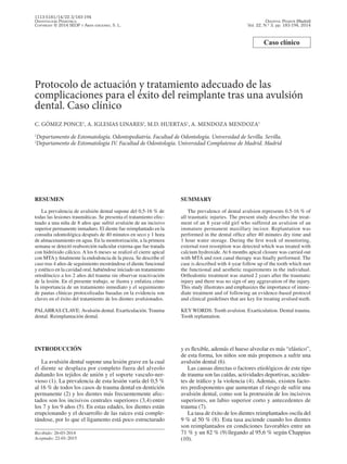
  • 1. Odontol Pediátr (Madrid) Vol. 22, N.º 3, pp. 183-194, 2014 1113-5181/14/22.3/183-194 Odontología Pediátrica Copyright © 2014 SEOP y Arán ediciones, S. L. Caso clínico Protocolo de actuación y tratamiento adecuado de las complicaciones para el éxito del reimplante tras una avulsión dental. Caso clínico C. Gómez Ponce1 , A. Iglesias Linares2 , M.D. Huertas1 , A. Mendoza Mendoza1 1 Departamento de Estomatología. Odontopediatría. Facultad de Odontología. Universidad de Sevilla. Sevilla. 2 Departamento de Estomatología IV. Facultad de Odontología. Universidad Complutense de Madrid. Madrid Recibido: 26-03-2014 Aceptado: 22-01-2015 Introducción La avulsión dental supone una lesión grave en la cual el diente se desplaza por completo fuera del alveolo dañando los tejidos de unión y el soporte vasculo-ner- vioso (1). La prevalencia de esta lesión varía del 0,5 % al 16 % de todos los casos de trauma dental en dentición permanente (2) y los dientes más frecuentemente afec- tados son los incisivos centrales superiores (3,4) entre los 7 y los 9 años (5). En estas edades, los dientes están erupcionando y el desarrollo de las raíces está comple- tándose, por lo que el ligamento está poco estructurado RESUMEN La prevalencia de avulsión dental supone del 0,5-16 % de todas las lesiones traumáticas. Se presenta el tratamiento efec- tuado a una niña de 8 años que sufrió avulsión de un incisivo superior permanente inmaduro. El diente fue reimplantado en la consulta odontológica después de 40 minutos en seco y 1 hora de almacenamiento en agua. En la monitorización, a la primera semana se detectó reabsorción radicular externa que fue tratada con hidróxido cálcico. A los 6 meses se realizó el cierre apical con MTAy finalmente la endodoncia de la pieza. Se describe el caso tras 4 años de seguimiento mostrándose el diente funcional y estético en la cavidad oral, habiéndose iniciado un tratamiento ortodóncico a los 2 años del trauma sin observar reactivación de la lesión. En el presente trabajo, se ilustra y enfatiza cómo la importancia de un tratamiento inmediato y el seguimiento de pautas clínicas protocolizadas basadas en la evidencia son claves en el éxito del tratamiento de los dientes avulsionados. Palabras clave: Avulsión dental. Exarticulación. Trauma dental. Reimplantación dental. SUMMARY The prevalence of dental avulsion represents 0.5-16 % of all traumatic injuries. The present study describes the treat- ment of an 8 year-old girl who suffered an avulsion of an immature permanent maxillary incisor. Replantation was performed in the dental office after 40 minutes dry time and 1 hour water storage. During the first week of monitoring, external root resorption was detected which was treated with calcium hydroxide. At 6 months apical closure was carried out with MTA and root canal therapy was finally performed. The case is described with 4-year follow up of the tooth which met the functional and aesthetic requirements in the individual. Orthodontic treatment was started 2 years after the traumatic injury and there was no sign of any aggravation of the injury. This study illustrates and emphasizes the importance of imme- diate treatment and of following an evidence-based protocol and clinical guidelines that are key for treating avulsed teeth. Key words: Tooth avulsion. Exarticulation. Dental trauma. Tooth replantation. y es flexible, además el hueso alveolar es más “elástico”, de esta forma, los niños son más propensos a sufrir una avulsión dental (6). Las causas directas o factores etiológicos de este tipo de trauma son las caídas, actividades deportivas, acciden- tes de tráfico y la violencia (4). Además, existen facto- res predisponentes que aumentan el riesgo de sufrir una avulsión dental, como son la protrusión de los incisivos superiores, un labio superior corto y antecedentes de trauma (7). La tasa de éxito de los dientes reimplantados oscila del 9 % al 50 % (8). Esta tasa asciende cuando los dientes son reimplantados en condiciones favorables entre un 71 % y un 82 % (9) llegando al 95,6 % según Chappius (10).
  • 2. 184 C. Gómez Ponce ET AL. Odontol Pediátr CASO CLÍNICO Descripción y diagnóstico Acude a la consulta de atención odontopediátrica especializada, una niña de 8 años de edad sin patología sistémica tras una caída en el colegio, a consecuencia de la cual sufrió la avulsión de un incisivo central superior derecho (Fig. 1A). Clínicamente se observaba formación de coágulo sanguíneo (Fig. 1B) y radiográficamente el alveolo vacío (Fig. 1C). El diente se mantuvo en una servilleta durante 40 minutos con inclusión posterior en agua durante una hora. Al llegar a la clínica el diente se mostraba íntegro observándose la no finalización en el desarrollo apical. Protocolo de tratamiento El diente fue reimplantado siguiendo las directrices actuales de la IADT (11). Se anestesió a la paciente (Mepivalem® 3 % SV, mepivacaína sin vasoconstrictor, Dentsply), se examinó el alveolo para comprobar que no existía obstáculo para el reimplante, se efectuó una radiografía oclusal y otra periapical con técnica de la bisectriz (aparato de rayos Sidexis), y se irrigó el alveolo con suero para eliminar el coágulo sanguíneo (Fig. 2). En este caso no se encontraron fracturas óseas pero se suturó una laceración en tejido blando a nivel del frenillo labial. Por otro lado, el diente se introdujo durante 5 minutos en una solución con 20 ml de suero fisiológico + 1 mg doxiciclina (Fig. 3) para favorecer la revascularización y la cicatrización periodontal (12-15). Posteriormente reimplantamos el diente con presión digital suave y aplicamos una férula flexible o semirrí- gida (Fig. 4A). Efectuamos una radiografía de control para verificar la posición correcta del diente (Fig. 4B). La férula (alambre 0,015 pulgadas tri-flex® ) se mantiene siete días (11,12,16,17). Se corroboró la existencia de protección inmunológica antitetánica, asimismo se prescribió cobertura antibiótica (50 mg/kg/día amoxicilina 7 días) (11,18). Siguiendo el protocolo internacional se sugiere como primera opción Fig. 2. Tras la anestesia se comprueba que no existe obstáculo para el reimplante y se irriga el alveolo con solución salina para eliminar el coágulo. Fig. 1. Aspecto clínico y radiológico inicial. A. Vista frontal del aspecto clínico inicial donde observamos ausencia del 11. B. Vista palatina del aspecto clínico inicial donde observamos el alveolo sangrante y vacío. C. Radiografía periapical de la zona para verificar la ausencia de fragmentos dentales en el alveolo o cuerpos extraños. Fig. 3. Introducción del diente en 20 ml de suero + 1 mg doxiciclina durante 5 minutos. A B C
  • 3. Vol. 22, N.º 3, 2014 Protocolo de actuación y tratamiento adecuado de las complicaciones para el éxito 185 del reimplante tras unaavulsión dental. Caso clínico la tetraciclina (doxiciclina 2 veces al día durante una semana con dosis adaptada para edad y peso) y como alternativa en pacientes jóvenes con riesgo de decolora- ción la amoxicilina o fenoximetilpenicilina (11,16). Por último, se dieron pautas de dieta blanda durante dos semanas e higiene con cepillo suave y enjuagues con clorhexidina (0,1 %) 2 veces al día durante una semana (11,12,16,19). Monitorización clínica y radiológica El trauma se sometió a un control clínico y radiográ- fico periódico a la semana, 4 semanas, 3 y 6 meses, al año, y luego anualmente durante cuatro años. La IADT recomienda un seguimiento anual durante cinco años (11,12,16). En el examen radiográfico realizado a la semana se observó una lesión por reabsorción radicular externa en el tercio medio de la raíz (Fig. 5A). Se extirpó la pul- pa, desbridando con limas K (Fig. 5B) e irrigando con hipoclorito al 5 % y se introdujo hidróxido cálcico puro hasta observar detención en la reabsorción radicular a los 6 meses (Fig. 5C). Posteriormente se facilitó el cie- rre apical mediante MTA (ProRoot® MTA, Dentsply) y se efectuó la endodoncia de la pieza con gutapercha y sellador (AH26, Dentsply) (Fig. 5D). El caso cuenta con un seguimiento anual durante 4 años (Figs. 6-8) obser- vando que el diente mantiene estabilidad, funcionalidad y estética aceptable durante el tiempo de monitorización. DISCUSIÓN El protocolo correcto ante una avulsión dental incluye unos primeros auxilios en el lugar del accidente: mante- ner la calma, manipular el diente a nivel de la corona y Fig. 4. Reimplante y ferulización. A. Vista clínica de la ferulización tras en reimplante. B. Verificación radiológica de la posición correcta tras el reimplante y la ferulización. Fig. 5. Seguimiento radiológico. A. En el seguimiento a la semana se detectó radiológicamente lesión por reabsorción radicular externa en el tercio medio de la raíz. B. Tratamiento intraconducto con hidróxido de calcio para frenar la reabsorción radicular externa. C. Detención en la reabsorción radicular a los 6 meses. D. Cierre apical mediante MTA y endodoncia. A B A C B D
  • 4. 186 C. Gómez Ponce ET AL. Odontol Pediátr asegurarnos de que se trata de un diente permanente ya que no es recomendable reimplantar dientes temporales (11,16). Si el diente estuviese sucio debemos lavarlo con agua o suero 10 segundos (11,16,21,22). Posteriormente el tratamiento de elección es su reim- plante (6,11,12,16,23). El reimplante inmediato (antes de 5 minutos) asegura la viabilidad del ligamento (6, 29-27)y una curación completa (23). Todos los esfuerzos deben hacerse para reimplantar el diente dentro de los primeros 15-20 minutos donde es posible la cicatrización periodontal (9,28-30). Si no fuese posible el diente debe almacenarse en un medio adecuado (11,12,16,23). El medio idóneo es la solución salina equilibrada de Hank (11,12,31-33). No obstante, la disponibilidad de este tipo de medio en el público general es irreal, con lo cual la leche desnatada, el suero o en último término la saliva pueden ser medios sustitutivos bastante útiles (1,11,16). El agua, utilizada en este caso, se debe evitar (11,16,34). Nuestro tratamiento y el pronóstico del diente es con- dicionado por 2 factores clínicos, el desarrollo apical y la condición de las células del ligamento (1,10-12,16,23). Esta última depende a su vez del tiempo en seco y del medio de almacenamiento (6,11,12,16,35,36). Fig. 6. Aspecto clínico y radiológico al año. Fig. 7. Aspecto clínico y radiológico a los 2 años. A A B B Respecto al desarrollo apical es importante cono- cer que sólo en dientes inmaduros es posible esperar la revascularización pulpar, por lo que sólo en estos casos no efectuaremos el tratamiento de conductos –salvo que existan indicios de necrosis pulpar (11,16) o se exce- dan los 60 minutos en seco (1,11,16). Para favorecer la revascularización se introduce el diente en doxiciclina o minociclina 0,05 % (1 mg/20 ml) 5 minutos antes del reimplante (11-16). En dientes con desarrollo apical maduro siempre efectuaremos tratamiento de conductos. El momento del tratamiento de conductos depende del periodo extraoral (11,12,16,37) y el hidróxido de calcio es el medicamento de elección (11). La viabilidad del ligamento es el otro factor a con- siderar. En este caso, estaba disminuida puesto que a los 30 minutos en seco se produce la muerte del 50 % de las células y a la hora apenas quedan células viables (38). Para aumentar la cicatrización periodontal debemos introducir el diente antes del reimplante en 1 mg minoci- clina o doxiciclina/20 ml solución salina 5 minutos. Sin embargo, cuando el tiempo extraoral en seco es superior a 60 minutos las células están necróticas y son invia- bles (23,28,30), debemos entonces eliminar el ligamento (11,12,16,23,39) y esperar la reabsorción sustitutiva y
  • 5. Vol. 22, N.º 3, 2014 Protocolo de actuación y tratamiento adecuado de las complicaciones para el éxito 187 del reimplante tras unaavulsión dental. Caso clínico anquilosis (11,16). El objetivo del reimplante en estos casos es estético, funcional, por razones psicológicas y para mantener el hueso alveolar (11,16,39). Para reducir la velocidad de reemplazo óseopueden introducirse estos dientes antes de su reimplantación en una solución al 2 % de fluoruro de sodio 20 minutos (11). La condición de las células del ligamento también pudo verse afectada por el medio de almacenamiento. El agua tiene baja osmolaridad (28,40), al ser hipotóni- ca provoca la lisis celular (41) y el aumento de la infla- mación en el reimplante (34). Según Andreasen (6) el almacenamiento en agua más de 20 minutos siempre dará como resultado reabsorción radicular, pero este mismo autor mostró cómo dientes reimplantados después de largos periodos extraorales no mostraron reabsorción radicular (44); también mostró un caso en el que dos incisivos que habían estado fuera de la boca 16 horas permanecieron funcionales 2 años. Shiu-yin Cho (45) mostró funcionalidad y estética durante 2 años de un diente que estuvo en seco 18 horas. Khalilak (46)descri- bió estabilidad durante 5 años de un diente reimplantado tras 270 minutos. Siguiendo estas recomendaciones el diente fue reimplantado después de una hora y cuarenta minutos extraorales considerando comprometida la via- bilidad del ligamento pero corroborando tras 4 años de seguimiento que el diente permanece funcional y estético en la cavidad oral. Adicionalmente, el caso descrito aborda una pieza dental inmadura donde había que sopesar el intento de revascularización frente a la posible aparición de com- plicaciones debido al largo periodo extraoral. Dos factores son críticos para que la revascularización pueda tener éxito, el tiempo (26,47) y la presencia de bacterias (26,47,48), ambos factores comprometidos en nuestro caso. Respecto al tiempo Kling (47)y Andreasen (26) consideran posible la revascularización si el dien- te se reimplanta dentro de los 45 minutos posteriores al trauma. Cvek (48) y otros autores (26,47) describen que la ocurrencia de revascularización es altamente dependiente de la presencia o ausencia de bacterias en la pulpa (13,15). En la actuación de dicho caso se intentó favorecer la revascularización introduciendo el diente en 20 ml de solución salina y 1 mg de doxiciclina (11). No consiguió la revascularización en este caso, pero pudo contribuir a la atenuación de la reabsorción radicular y a la curación periodontal. Además, el tratamiento sisté- mico antibiótico también ayudó a evitar la proliferación bacteriana y limitar la reabsorción inflamatoria (49,50). En el control radiológico a la semana se detectó reab- sorción radicular inflamatoria externa en el tercio medio de la raíz. Este tipo de reabsorción está asociada con necrosis e infección de la pulpa, de forma que las bacterias entran en el espacio periodontal a través de los túbulos dentina- rios (4) y junto al daño en el cemento resulta en reabsor- ción inflamatoria externa (51). Actualmente el tratamiento, prevención o limitación de la reabsorción radicular incluye la terapia con hidróxido de calcio (52), aunque también existen buenos resultados con una combinación de antibió- tico y corticoide como Ledermix (53-56). Estudios actua- les plantean el posible uso de corticoides (57), calcitonina (58), acetazolamida (59), bifosfonatos (60,61)o derivado de la matriz del esmalte (62,63). Sin embargo, hoy día, el hidróxido cálcico sigue siendo el medicamento de elección y fue el que usamos en el presente caso. Posteriormente se facilitó el cierre apical mediante MTA y finalmente se efectuó la endodoncia de la pieza. CONCLUSIONES Reimplantar el diente a la mayor brevedad posible o mantenerlo en un medio adecuado son parámetros clí- nicos fundamentales en el pronóstico de la pieza avul- sionada. Fig. 8. A. Aspecto radiológico a los 3 años y medio durante el movimiento ortodóncico. B. Seguimiento radiológico a los 4 años. El diente se mantuvo estable funcional y estéticamente aceptable. No se observó reabsorción inflamatoria ni anquilosis. A B
  • 6. 188 C. Gómez Ponce ET AL. Odontol Pediátr El estado del ápice y el periodo extraoral en seco condicionan dramáticamente el pronóstico del diente avulsionado. El seguimiento de pautas clínicas protocolizadas basa- das en la evidencia determinan en gran medida el éxito del tratamiento a pesar de su mal pronóstico inicial. BIBLIOGRAFÍA 1. Trope M. Clinical management of the avulsed tooth: present strategies and future directions. Dent Traumatol 2002;18:1-11. 2. Davidovich E, Moskovitz M, Moshonov J. Replantation of an immature permanent central incisor following preeruptive trau- matic avulsion. Dent Traumatol 2008;24(5):e47-52. 3. Gonda F, Nagase M, Chem RB, Yakata H, Nakajima T. Replan- tation: An analysis of 29 teeth. Oral Surg Oral Med Oral Pathol 1990;70:650-5. 4. Tzigkounakis V, Merglová V, Hecová H, Netolický J. Retrospec- tive clinical study of 90 avulsed permanent teeth in 58 children. Dent Traumatol 2008;24:598-602. 5. Baginska J, Wilczynska-Borawska M. First-aid algorithms in dental avulsion. J Sch Nurs 2012;28(2):90-4. 6. Andreasen JO, Borum, MK, Jacobsen HL, Andreasen FM. Replantation of 400 avulsed permanent incisors. 4. Factors relat- ed to periodontal ligament healing. Endodontics Dent Traumatol 1995;11:76-89. 7. Brin I, Ben-Bassat Y, Heling I, Brezniak N. Profile of an ortho- dontic patient at risk of dental trauma. Endod Dent Traumatol 2000;16:111-5. 8. Steiner DR. Avulsed maxillary central incisors: The case for replantation. Am J Orthod Dentofacial Orthop 2012;142(1):8, 10, 12, 14, 16. 9. Andersson L, Bodin I. Avulsed human teeth replanted within 15 minutes —a long-term clinical follow-up study. Endod Dent Traumatol 1990;6:37-42. 10. Chappuis V, von Arx T. Replantation of 45 avulsed permanent teeth: A 1-year follow-up study. Dent Traumatol 2005;21(5): 289-96. 11. Andersson L, Andreasen JO, Day P, Heithersay G, Trope M, Diangelis AJ, et al. International Association of Dental Trau- matology guidelines for the management of traumatic den- tal injuries: 2. Avulsion of permanent teeth. Dent Traumatol 2012;28(2):88-96. 12. Flores MT, Andreasen JO, Bakland LK, Feiglin B, Gutmann JL, Oikarinen K et al.; International Association of Dental Trauma- tology. Guidelines for the evaluation and management of trau- matic dental injuries. Dent Traumatol 2001;17:193-8. 13. Cvek M, Cleaton-Jones P, Austin J, Lownie J, Kling M, Fatti P. Effect of topical application of doxycycline on pulp revascular- ization and periodontal healing in reimplanted monkey incisors. Endod Dent Traumatol 1990;6(4):170-6. 14. Yanpiset K, Trope M. Pulp revascularization of replanted imma- ture dog teeth after different treatment methods. Endod Dent Traumatol 2000;16:211-7. 15. Ritter AL, Ritter AV, Murrah V, Sigurdsson A, Trope M. Pulp revascularization of replanted immature dog teeth after treat- ment with minocycline and doxycycline assessed by laser Dop- pler flowmetry, radiography, and histology. Dent Traumatol 2004;20:75-84. 16. Flores MT,Andersson L,Andreasen JO, Bakland LK, Malmgren B, Barnett F. Guidelines for the management of traumatic den- tal injuries. II. Avulsion of permanent teeth. Dent Traumatol 2007;23:130-6. 17. Andreasen JO, Friskopp J, Blomloff L. Fiberglass splinting of traumatized teeth. J Dent Child 1983;3:21-24. 18. Hammarstrom L, Blomloff L, Feiglin B, Andersson L, Lindskog S. Replantation of teeth and antibiotic treatment. Endod Dent Traumatol 1986;2:51-7. 19. Ram D, Cohenca N. Therapeutic protocols for avulsed permanent teeth: Review and clinical update. Pediatr Dent 2004;26:251-5. 20. Davidovich E, Moskovitz M, Moshonov J. Replantation of an immature permanent central incisor following pre-eruptive trau- matic avulsion. Dent Traumatol 2008;24:e47-e52. 21. Nesiama JO, Sinn DP. Tooth avulsion. Clinical Pediatric Emer- gency Medicine 2010;11:108-11. 22. Emerich K, Kaczmarek J. First aid for dental trauma caused by sports activities. State of knowledge, treatment and prevention. Sports Med 2010;40(5):361-6. 23. Trope M. Avulsion of permanent teeth: Theory to practice. Den- tal Traumatology 2011;27(4):281-94. 24. Andreasen JO, Borum MK, Jacobsen HL, Andreasen FM. Replantation of 400 avulsed permanent incisors. 1. Diagnosis of healing complications. Endod Dent Traumatol 1995;11:51-8. 25. Andreasen JO, Borum MK, Jacobsen HL, Andreasen FM. Replantation of 400 avulsed permanent incisors. 2. Factors related to pulpal healing. Endod Dent Traumatol 1995;11:59-68. 26. Andreasen JO, Borum MK, Jacobsen HL, Andreasen FM. Replantation of 400 avulsed permanent incisors. 3. Factors related to root growth. Endod Dent Traumatol 1995;11:69-75. 27. Kinirons MJ, Gregg TA, Welbury RR, Cole BOI. Variations in the presenting and treatment features in reimplanted permanent incisors in children and their effect on the prevalence of root resorption. Br Dent J 2000;189:263-6. 28. Andreasen JO. The effect of extra-alveolar period and stor- age media upon periodontal and pulpal healing after replanta- tion of mature permanent incisors in monkeys.Int J Oral Surg 1981;10:43-53. 29. Barrett EJ, Kenny DJ. Avulsed permanent teeth: A review of the literature and treatment guidelines. Endod Dent Traumatol 1997;13:153-63. 30. Soder PO, Otteskog P, Andreasen JO, Modeer T. Effect of dry- ing on viability of periodontal membrane. Scand J Dent Res 1977;85:167-72. 31. Hiltz J, Trope M. Vitality of human lip fibroblasts in milk. Hank’s balanced salt solution and Via Span storage media. Endod Dent Traumatol 1991;7:69-72. 32. Ashkenazi M, Sarnat H, Keila S. In vitro viability, mitogenic- ity and clonogenic capacity of periodontal ligament cells after storage in six different media. Endod Dent Traumatol 1999;15: 149-56. 33. Trope M, Friedman S. Periodontal healing of replanted dog teeth stored in Viaspan, milk and Hanks Balanced Salt Solution. Endod Dent Traumatol 1992; 8:183-8. 34. Blomlof L. Milk and saliva as possible storage media for trau- matically exarticulated teeth prior to replantation. Swed Dent J Suppl 1981;8:1-26. 35. Pettiette M, Hupp J, Mesaros S, Trope M. Periodontal healing of extracted dogs’teeth air-dried for extended periods and soaked in various media. Endod Dent Traumatol 1997;13:113-8. 36. Lin DG, Kenny DJ, Barrett EJ, Lekic P, McCulloch CAG. Stor- age conditions of avulsed teeth affect the phenotype of cultured human periodontal ligament cells. J Periodont Res 2000;35: 42-50. 37. Giannetti L, Murri A. Clinical evidence and literature to com- pare two different therapeutic protocols in tooth avulsion. Eur J Pediatr Dent 2006;7:122-30. 38. Flogia I. Avulsión dentaria. Otros recursos terapéuticos. EJER 2012;11(1):575-96. 39. Panzarini SR, Gulinelli JL, Poi WR, Sonoda CK, D Pedrini, Brandini DA. Treatment of root surface in delayed tooth replan- tation:Areview of literature. Dent Traumatol 2008;24(3):277-82. 40. Sigalas E, Regan JD, Kramer PR, Witherspoon DE, Opperman LA. Survival of human periodontal ligament cells in media pro- posed for transport of avulsed teeth. Dent Traumatol 2004;20:21-8. 41. Marino TG, West LA, Liewehr FR, Mailhot JM, Buxton TB, Runner RR, et al. Determination of periodontal ligament cell viability in long shelf-life milk. J Endod 2000;26:699-702. Correspondencia: Cristina Gómez Ponce Departamento de Estomatología Odontopediatría Facultad de Odontología Universidad de Sevilla e-mail: cristinagomezponce@hotmail.com
  • 7. Vol. 22, N.º 3, 2014 Protocol of action and appropriate treatment of complications for replantation 189 success following dental avulsion. A case report Protocol of action and appropriate treatment of complications for replantation success following dental avulsion. A case report C. Gómez Ponce1 , A. Iglesias Linares2 , M.D. Huertas1 , A. Mendoza Mendoza1 1 Department of Dentistry. Pediatric Dentistry. Faculty of Dentistry. University of Seville. Seville, Spain. 2 Department of Dentistry IV. Faculty of Dentistry. Universidad Complutense de Madrid. Madrid, Spain Clinical case 42. Pohl Y, Tekin U, Boll M, FilippiA, Kirschner H. Investigation on a cell culture medium for storage and transportation of avulsed teeth. Aust Endod J 1999;25:70-5. 43. Andersson L, Bodin I, Sorensen S. Progression of root resorption following replantation of human teeth after extended extraoral storage. Endod Dent Traumatol 1989;5:38-47. 44. Andreasen JO, Hjorting-Hansen E. Replantation of teeth. I. Radiographic and clinical study of 110 human teeth replanted after accidental loss. Acta Odontol Stand 1966;24:263-86. 45. Cho SY, Cheng AC. Replantation of an avulsed incisor after prolonged dry storage: A case report. J Can Dent Assoc 2002;68(5):297-300. 46. Khalilak Z, Shikholislami M, Mohajeri L. Delayed tooth replan- tation after traumatic avulsion: A case report. Iran Endod J 2008;3(3):86-9. 47. Kling M, Cvek M, Mejare I. Rate and predictability of pulp revascularization in therapeutically reimplanted permanent inci- sors. Endod Dent Traumatol 1986;2(3):83-9. 48. Cvek M, Cleaton-Jones P, Austin J, Lownie J, Kling M, Fatti P. Pulp revascularization in reimplanted immature monkey incisors – predictability and the effect of antibiotic systemic prophylaxis. Endod Dent Traumatol 1990;6:157-69. 49. Hammarstrom L, Pierce A, Blomlof L, Feiglin B, Lindskog S. Tooth avulsion and replantation: A review. Endod Dent Trau- matol 1986;2:1-9. 50. Sae-Lim V, Wang CY, Trope M. Effect of systemic tetracicline and amoxicillin on inflammatory root resorption of replanted dog, teeth. Endod Dent Traumatol 1998;14:216-20. 51. Tronstad L. Root resorption etiology, terminology and clinical manifestations.Endod Dent Traumatol 1988;4:241. 52. Barnett F. The role of endodontics in the treatment of luxated permanent teeth. Dent Traumatol 2002;18(2):47-56. 53. Bryson EC, Levin L, Banchs F, Abbott PV, Trope M. Effect of immediate intracanal placement of Ledermix Paste(R) on healing of replanted dog teeth after extended dry times. Dent Traumatol 2002;18:316-21. 54. Wong KS, Sae-Lim V. The effect of intracanalLedermix on root resorption of delayed-replanted monkey teeth. Dent Traumatol 2002;18:309-15. 55. Pierce A, Lindskog S. The effect of an antibiotic corticoste- roid combination on inflammatory root resorption. J Endod 1988;14:459-64. 56. Thong YL, Messer HH, Siar CH, Saw LH. Periodontal response to two intracanal medicaments in replanted monkey incisors. Dent Traumatol 2001;17:2549. 57. Kirakozova A, Teixeira FB, Curran AE, Gu F, Tawil PZ, Trope M. Effect of intracanal corticosteroids on healing of replanted dog teeth after extended dry times. J Endod 2009;35:663-7. 58. Pierce A, Berg JO, Lindskog S. Calcitonin as an alternative ther- apy in the treatment of root resorption. J Endod 1988;14:459-65. 59. Mori GG, Garcia RB. Microscopic study of the effect of rootsur- face treatment with acetazolamida in avulsed and reimplanted rat teeth. Rev Fac Odontol Bauru 2002;10:180-5. 60. Lustrosa-Pereira A, Garcia RB, de Moraes IG, Bernadineli N, Bramante CM, Bortoluzzi EA. Evaluation of the topical effect of alendronate on the root surface of extracted and replant- ed teeth. Microscopic analysis on rats’ teeth. Dent Traumatol 2006;22:30-5. 61. Levin L, Bryson EC, Caplan D, Trope M. Effect of topical alen- dronate on root resorption of dried replanted dog teeth. Dent Traumatol 2001;17:120-6. 62. Schjott M, Andreasen JO. Emdogain does not prevent progres- sive root resorption after replantation of avulsed teeth: A clinical study. Dent Traumatol 2005;21:46-50. 63. Fillippi A, Pohl Y, von Arx T. Treatment of replacement resorp- tion by intentional replantation, resection of the ankylosedsites, and Emdogain – results of a 6-year survey. Dent Traumatol 2006;22:307-11. RESUMEN La prevalencia de avulsión dental supone del 0,5-16 % de todas las lesiones traumáticas. Se presenta el tratamiento efec- tuado a una niña de 8 años que sufrió avulsión de un incisivo superior permanente inmaduro. El diente fue reimplantado en la consulta odontológica después de 40 minutos en seco y 1 hora de almacenamiento en agua. En la monitorización, a la primera semana se detectó reabsorción radicular externa que fue tratada con hidróxido cálcico. A los 6 meses se realizó el cierre apical con MTAy finalmente la endodoncia de la pieza. Se describe el caso tras 4 años de seguimiento mostrándose el diente funcional y estético en la cavidad oral, habiéndose iniciado un tratamiento ortodóncico a los 2 años del trauma sin observar reactivación de la lesión. En el presente trabajo, se ilustra y enfatiza cómo la importancia de un tratamiento inmediato y el seguimiento de pautas clínicas protocolizadas basadas en la evidencia son claves en el éxito del tratamiento de los dientes avulsionados. Palabras clave: Avulsión dental. Exarticulación. Trauma dental. Reimplantación dental. SUMMARY The prevalence of dental avulsion represents 0.5-16 % of all traumatic injuries. The present study describes the treat- ment of an 8 year-old girl who suffered an avulsion of an immature permanent maxillary incisor. Replantation was performed in the dental office after 40 minutes dry time and 1 hour water storage. During the first week of monitoring, external root resorption was detected which was treated with calcium hydroxide. At 6 months apical closure was carried out with MTA and root canal therapy was finally performed. The case is described with 4-year follow up of the tooth which met the functional and aesthetic requirements in the individual. Orthodontic treatment was started 2 years after the traumatic injury and there was no sign of any aggravation of the injury. This study illustrates and emphasizes the importance of imme- diate treatment and of following an evidence-based protocol and clinical guidelines that are key for treating avulsed teeth. Key words: Tooth avulsion. Exarticulation. Dental trauma. Tooth replantation.
  • 8. 190 C. GÓMEZ PONCE ET AL. Odontol Pediátr INTRODUCTION Tooth avulsion is a serious injury during which a tooth is completely displaced from the alveolus, attachment damage occurs and the neurovascular bundle is affected (1). The prevalence of this lesion varies between 0.5 % and 16 % of all dental trauma cases related to the perma- nent dentition (2) and the teeth that are more commonly affected are the upper central incisors (3,4) between the ages of 7 and 9 years (5).At this age the teeth have erupted but root development is not quite complete. The periodon- tal ligament lacks some structure and it is flexible and, in addition, the alveolar bone is more “elastic”. Because of this children are more inclined to suffer the avulsion of a tooth (6). The direct causes or etiological factors behind this type of traumatic injury are falls, sporting activities, traffic accidents and violence (4). In addition there are pre- disposing factors that increase the risk of suffering tooth avulsion such as protrusion of the upper incisors, short lower lip, and antecedents of trauma (7). The success rate of replanted teeth ranges between 9 % and 50 % (8). This rate increases when the teeth are replanted in favorable conditions to 71 % and 82 % (9) even reaching 95.6 % according to Chappius (10). CASE REPORT Description and diagnosis An 8 year-old girl, with no systemic disease, attended the specialized pediatric dentistry department after a fall at school involving the avulsion of the upper right central incisor (Fig. 1A). The clinical examination revealed the formation of a blood clot (Fig. 1B) and the radiographic findings showed an empty alveolus (Fig. 1C). The tooth had been kept in a napkin for 40 minutes and it was later placed in water for an hour. On arrival at the clinic the tooth was intact and incomplete apical closure could be observed. Treatment protocol The tooth was reimplanted following the current guidelines of the IADT (11). The patient was anesthe- tized with (3 % Mepivalem® SV, mepivacaine without vasoconstrictor, Dentsply). The alveolus was examined in order to verify that no obstacles would be encountered during the replantation and an occlusal radiograph and another periapical radiograph were carried out using the bisecting angle technique (Sidexis X-ray machine). The alveolus was irrigated with saline in order to eliminate the blood clot (Fig. 2). Bone fractures were not found but a laceration of the soft tissues by the labial frenulum was sutured. The tooth was placed for 5 minutes in a solu- tion of 20 ml of saline with 1 mg of doxycycline (Fig. 3) in order to encourage revascularization and periodontal healing (12-15). The tooth was then reimplanted with gentle finger pressure and a flexible or semi-rigid splint was fitted (Fig. 4A). A control radiograph was taken in order to verify the correct position of the tooth (Fig. 4B). The Fig. 2. Following anesthesia, the area was checked to ensure there was nothing hampering the replantation. The alveolar socket was irrigated with saline solution in order to eliminate the blood clot. Fig. 1. Initial clinical and radiologic findings. A. Frontal view of initial clinical appearance showing missing 11; B. Palatal view of initial clinical appearance showing bleeding alveolar socket. C. Periapical radiograph of the area in order to ensure no tooth fragment or foreign body in the alveolar socket. A B C
  • 9. Vol. 22, N.º 3, 2014 Protocol of action and appropriate treatment of complications for replantation 191 success following dental avulsion. A case report splint (0.015 inch tri-flex® wire) was kept in place for a week (11,12,16,17). Her anti-tetanus status was checked, and antibiotic was prescribed (50 mg/kg/day amoxicillin for a week) (11,18). The international protocol suggests tetracycline as a first option (doxycycline twice a day for a week with the dosage adapted to age and weight) and for young patients at risk of discoloration amoxicillin or phenoxy- methylpenicillin (11,16). Lastly, instructions were given regarding a soft diet for two weeks and hygiene with a soft toothbrush and chlorhexidine rinses (0.1 %) twice a day for a week (11,12,16,19). Clinical monitoring and radiology The traumatic injury was monitored clinically and radiographically over regular periods: At one week, 4 weeks, 3 and 6 months and at one year, and then annu- ally for four years. The IADT recommends an annual follow-up of five years (11,12,16). During the radiographic examination a week later a lesion was observed as a result of external root resorp- tion in the mid-third of the root (Fig. 5A). The pulp was extracted, debridement was performed with a K-file (Fig. 5B) and it was irrigated with 5 % hypochlorite. Pure calcium hydroxide was introduced until the root resorption was arrested at 6 months (Fig. 5C). Apical closure was then achieved with MTA (Pro- Root® MTA, Dentsply) and root canal treatment was car- ried out with gutta-percha and a sealant (AH26, Dents ply) (Fig. 5D). The case has had an annual follow-up of 4 years (Figs. 6-8) and the tooth has shown stability and functionality during monitoring, and an acceptable aesthetic appearance. DISCUSSION The correct protocol for an avulsed tooth includes first aid at the site of the accident: Keeping calm, handling the tooth by the crown and ensuring that it is a permanent tooth, as the replantation of primary teeth is not recom- mendable (11,16). If the tooth is dirty it should be cleaned with water or saline for 10 seconds (11,16,21,22). After this the treatment of choice is replantation (6,11,12,16,23). Immediate replantation (after 5 minutes) ensures the viability of the ligament (6,29-27) and complete healing (23). Every effort should be made to reimplant the tooth within the first 15-20 minutes to facilitate periodontal healing (9,28-30). If not possible the tooth should be stored in a suitable media (11,12,16,23). The ideal medium is Hanks balanced salt solution (11,12,31-33). Nevertheless, the availability of this type of medium among the general public is unrealistic, and skimmed milk, saline or as a last resort saliva can be quite useful substitutes (1,11,16). Water in this case should be avoided (11,16,34). The treatment given and the prognosis of the tooth is conditioned by two clinical factors, root development stage and the condition of the ligament cells (1,10-12,16,23). The latter depends on the dry time and the storage medium (6,11,12,16,35,36). Fig. 3. The tooth was placed in 20 ml of saline solution + 1 mg doxycycline for 5 minutes. Fig. 4. Replantation and splinting. A. Clinical appearance of splinting after the replantation. B. correct position after replantation and splinting is verified radiographically. A B
  • 10. 192 C. GÓMEZ PONCE ET AL. Odontol Pediátr With regard to root development stage, it is important to be aware that with immature teeth it is possible to wait for pulp revascularization, and only in these cases should root canal therapy not be carried out (unless there is evidence of pulp necrosis (11,16) or if there is dry time of over 60 minutes (1,11,16). In order to promote revas- cularization, the tooth should be introduced in doxycy- cline or minocycline 0.05 % (1 mg/20 ml) for 5 minutes before replantation (11-16). Root canal therapy should always be carried out in teeth with mature apexes. The extraoral period (11,12,16,37) will determine when root canal therapy is carried out, and calcium hydroxide is the treatment of choice (11). The viability of the ligament is another factor to take into consideration. In the present case it was reduced because after 30 minutes dry time, 50 % of the cells die and after an hour there will hardly be any viable cells at all (38). In order to encourage periodontal healing, the tooth should be introduced before the replantation into 1 mg of minocycline or doxycycline/20 ml of saline solution for 5 minutes. However, when the extraoral dry time is over 60 minutes the cells will be necrotic and non- viable (23,28,30), and the ligament should be eliminated (11,12,16,23,39) as replacement resorption and ankylosis will ensue (11,16). The aim of the replantation in these cases is aesthetic and functional, due to psychological reasons and to preserve the alveolar bone (11,16,39). In order to reduce the rate of bone replacement, these teeth can be placed in a solution of 2 % sodium fluoride for 20 minutes (11). The condition of the ligament cells may also have been affected by the storage medium. Water has low osmolarity (28,40), and the hypotonic effect will lead to cell lysis (41) and increased inflammation of the replan- tation (34). According to Andreasen (6) water storage of more than 20 minutes will always lead to root resorption, but this same author showed how replanted teeth after a extended extraoral periods did not result in induced root resorption (44). He also reported a case in which two Fig. 5. Radiological follow-up. A. During follow-up at one week, the lesion was detected radiologically due to external root resorption in the mid-third of the root. B. Root canal treatment with calcium hydroxide in order to arrest external root resorption. C. Arrested root resorption at 6 months. D. Closure of the apex using MTA and root canal therapy. A C B D Fig. 6. Clinical and radiographic appearance at one year. A B
  • 11. Vol. 22, N.º 3, 2014 Protocol of action and appropriate treatment of complications for replantation 193 success following dental avulsion. A case report Fig. 7. Clinical and radiographic appearance at two years. incisors that had been outside the mouth for 16 hours remained functional for two years. Shiu-yin Cho (45) demonstrated the functionality and aesthetic appearance for 2 years of a tooth that had been kept dry for 18 hours. Khalilak (46) described stability over 5 years of a tooth that was replanted after 270 minutes. Following these recommendations the tooth was replanted after one hour and forty minutes extraoral time although the viability of the ligament was considered compromised but, after a follow-up of four years the tooth showed functional stability and it had an acceptable aesthetic appearance in the oral cavity. Additionally, the case described concerned an imma- ture tooth where the attempt to revascularize had to be weighed up against the possible appearance of complica- tions due to the extended extraoral time. Two factors are critical for revascularization to be successful, time (26,47) and the presence of bacteria (26,47,48), and both factors but our casein jeopardy. With regard to time, Kling (47) and Andreasen (26) consider that revascularization is possible if the tooth is replanted within 45 minutes of the trauma. Cvek (48) and other authors (26,47) showed that pulp revasculariza- tion is highly dependent upon the presence or absence of bacteria in the pulp (13,15). In this case of ours, revascu- larization was encouraged by placing the tooth in 20ml of saline solution and 1 mg of doxicyline (11). Revascu- larization was not achieved in this case but it may have contributed to the attenuation of root resorption and peri- odontal healing. In addition systemic antibiotic treatment also helped to avoid bacterial proliferation and to limit inflammatory root resorption (49,50). Monitoring by X-ray a week later revealed external inflammatory root resorption at the mid-third of the root. This type of resorption is associated with necrosis and pulp infection, as bacteria enter the periodontal space through the dentinal tubules (4) and, with the damage to the cementum, Fig. 8. A. Radiographic appearance at 3 years and a half during orthodontic movement. B. Radiological follow-up at 4 years. The tooth remained stable and functional and it had an aesthetically acceptable appearance. There was no inflammatory resorption or ankylosis. A B A B
  • 12. 194 C. GÓMEZ PONCE ET AL. Odontol Pediátr there is external inflammatory resorption (51). Currently the treatment, prevention or control of root resorption includes calcium hydroxide therapy (52), although there are also good results with a combination of antibiotics and corti- costeroid such as Ledermix (53-56). Current studies raise the possibility of using corticosteroids (57), calcitonine (58), acetazolamide (59), bisphosphonates (60,61) or enamel matrix derivatives (62,63). However, calcium hydroxide continues being the medication of choice and it was used in this case study. Closure of the root apex was carried out with MTA and lastly root canal therapy was performed. CONCLUSIONS Replantation of the tooth as quickly as possible, or maintaining it in suitable media, are basic clinical param- eters for the prognosis of an avulsed tooth. The condition of the apex and the extraoral dry time have a dramatic effect on the prognosis of the avulsed tooth. Following evidence-based clinical guidelines that are protocolized will determine to a large extent the success of the treatment despite the initial prognosis being poor.