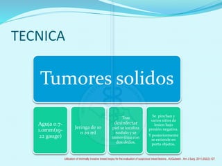
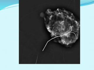
DR. GILBERTO J. SOLANO APARICIO
R6 ONCOGINECOLOGIA
IECAN
BIOPSIAS
 Muestra de tejido o células que se obtiene de un tumor,
indicada para diagnostico definitivo y preciso de una
neoplasia.
 Clasificación: Insicionales, Excisionales, Por punción,
Por aspiracion.
Utilization of minimally invasive breast biopsy for the evaluation of suspicious breast lesions., AUGutwein , Am J Surg. 2011;202(2):127.
Diagnostico de lesiones de la mama
Utilization of minimally invasive breast biopsy for the evaluation of suspicious breast lesions., AUGutwein , Am J Surg. 2011;202(2):127.
Posterior a diagnostico por la
exploracion clinica y tecnicas de imagen
, la diferentes variables de lesiones en
la mama se estudian según las
caracteristicas individuales.
ADENOPATIAS
LESIONES NO PALPABLES SOSPECHOSAS DE
MALIGNIDAD CON TECNICAS DE IMAGEN
SECRECIONES,
LESIONES ECCEMATOSAS.
INFLAMACION DE LA MAMA
QUISTES
TUMOR SOLIDO PB
BENIGNO
TUMOR SOLIDO PB
MALIGNO
Diagnostico de lesiones de la mama
Utilization of minimally invasive breast biopsy for the evaluation of suspicious breast lesions., AUGutwein , Am J Surg. 2011;202(2):127.
Diagnostico de lesiones de la mama
Utilization of minimally invasive breast biopsy for the evaluation of suspicious breast lesions., AUGutwein , Am J Surg. 2011;202(2):127.
BIOPSIAS
BAAF
AGUJA DE
CORTE
VACUUM
BX
QUIRÚRGICA
Especificidad : 97%
Sensibilidad : 72-94%
Especificidad: 99%
Sensibilidad: 91%
Similar a aguja de
corte
Alta sensibilidad
100%
FN: 0-32% Lesiones sólidos FN: 0.5 a 22% FN: 1.0%
Operador
dependiente
No diferencia
invasión
Complicaciones:
Múltiples punciones
Lesión de músculos,
intratorácica o
periductal
Mayor sangrado
Riesgo de hematoma
Mayor volumen de
tejido
Fallas de 1.1%
Recomendada para
evaluación de
ganglios axilares
Quistes
DX e IHQ Mejores resultados
Biopsia de
calcificaciones
Costo
Anestesia.
cicatriz
Calibre de aguja:
22-25 g
11-14 g 9, 11, 14 g
-
Utilization of minimally invasive breast biopsy for the evaluation of suspicious breast lesions., AUGutwein , Am J Surg. 2011;202(2):127.
HISTORIA
CLÍNICA
IMAGEN
EXPLORACIÓ
N FÍSICA
HISTOPATOLÓGICO
Cancer. 2001;91(7):1231
Exploración físicaObjetivo: determinar lesión dominante, tamaño y localización
- 4 % se detecta por este método.
- Sensibilidad muy Baja.
- 4 % cuando el tumor es de 0.3
cms.
- > 90 % en tumores de > de 1 cms.
- Habilidad del experto : 43 %
Bland K, Copeland E. The Breast. 2009.
imagen
MASTOGRAFÍA
-80% lesiones no palpables
- S: 85 % E: 87-99.5%
- FP: 3.6 -77.4%
ULTRASONIDO MAMARIO Y AXILAR
- Estudio complementario
- S: 55-85% E: 55-79%
RESONANCIA MAGNÉTICA
- Estudio complementario
- S: 91-100% E: 37-67% - FP: 2.6-79.7%
TOMOSÍNTESIS
- Proyecciones Rayos X en 3D
- S: 89% E: 92%
Eur Radiol. 2012 Jan;22(1):26-38
BIRADS
Breast Imaging Reporting and Data System
Categoría Descripción Porcentaje Recomendación
0 Estudio insuficiente - Necesita otros estudios
1 estudio normal 0% control habitual
2 Patología Benigna 0% Control habitual
3 Sugestiva de benignidad < 2% Control 6 meses
4 a
4 b
4 c
Baja a moderada sospecha
Moderada sospecha
Moderada a alta sospecha
> 2 al 10%
11 al 40%
41 al 94%
Considerar estudio
histológico
5 Alta sospecha malignidad 95% Estudio histológico
6 Malignidad confirmada 100% -
American College of Radiology, Reston, VA, 2003.
Aspiración con
aguja fina
Aspiracion de
liquidos de quiste o
material celular de un
tumor solido.
indiaciones
Tumores
aparentemente
quisticos.
Tumores solidos
bien definidos,
mayores de 1cm
Carcinomas
inoperables(
confirmacion dx)
Exclusion de
cancer en paciente
con inflamacion.
Ganglios
sospechosos
Utilization of minimally invasive breast biopsy for the evaluation of suspicious breast lesions., AUGutwein , Am J Surg. 2011;202(2):127.
TECNICA
Tumores solidos
Aguja 0.7-
1.0mm(19-
22 gauge)
Jeringa de 10
o 20 ml
Tras
desinfectar
piel se localiza
nodulo y se
inmoviliza con
dos dedos.
Se pinchan y
varios sitios de
lesion bajo
presión negativa.
Y posteriormente
se extiende en
porta objetos.
Utilization of minimally invasive breast biopsy for the evaluation of suspicious breast lesions., AUGutwein , Am J Surg. 2011;202(2):127.
TECNICA
Quistes
Vaciado
completo.
Llenado con aire y
hacer estudio
ecográfico o mamo
grafico
3m resolución
10 a 20%
desarrollan
nuevos quistes..
Utilization of minimally invasive breast biopsy for the evaluation of suspicious breast lesions., AUGutwein , Am J Surg. 2011;202(2):127.
Malignidad 1 por 100 000.
1% carcinomas quisticos.
Postmenopausica y el contenido de aspirado suele ser hematico
Marchan 1993;Moore 1991
Utilization of minimally invasive breast biopsy for the evaluation of suspicious breast lesions., AUGutwein , Am J Surg. 2011;202(2):127.
BAAFINSTRUMENTOS TÉCNICA
 Solución antiséptica *
isodine
 Jeringa de 10 ml
 Aguja de 22-25 g
 Portaobjetos
 Alcohol 90 %
 Preparación adecuada para
la piel
 Mano no dominante
estabiliza la lesión o GL.
 Introducir aguja
 Succión repetida
 Fijación en porta objetos
American College of Obstetricians and Gynecologists: Breast con- cerns in the
adolescent. ACOG Committee Opinion 350. Obstet Gynecol 2006;108:1329.
BAAF
 MALIGNIDAD:
 quistes
 1% (1-2/100).
 Carcinomas
quísticos.
American College of Obstetricians and Gynecologists: Breast con- cerns in the
adolescent. ACOG Committee Opinion 350. Obstet Gynecol 2006;108:1329.
BIOPSIA CON AGUJA (TROCAR)
 Presentado por primera
vez en 1989 por Parker y
cols.
 Está basado en el muestreo
de una lesión mediante el
uso de un dispositivo
manual, que contiene un
sistema de corte gatillado
automáticamente al pre-
sionar un botón
 permite así cortar
cilindros de tejido, de
grosor variable según
la cánula que se use,
específicamente en
mama 14 o 12 Gauge, y
de longitud hasta 2,2
cm
Utilization of minimally invasive breast biopsy for the evaluation of suspicious breast lesions., AUGutwein , Am J Surg. 2011;202(2):127.
BIOPSIA CON AGUJA (TROCAR)
Tumores cercanos a la piel
Bien definidos.
Palpables.
No menores de
2cm.
Anestesia local.
 BAAF Identifica 67-
89% de canceres.
 Trocar: entre el 75 y
el 99%
Utilization of minimally invasive breast biopsy for the evaluation of suspicious breast lesions., AUGutwein , Am J Surg. 2011;202(2):127.
BIOPSIA TRUCUT
Wells S. atlas of breast surgery. edición única. Washington DC: Mosby; 1998
Smith: Netter's Obstetrics and Gynecology, 2nd ed, 2008
• Preparación de la piel
• Anestésico local
• Pequeña incisión en piel con
bisturí # 11
• Estabilidad de la lesión con la
mano no dominante.
• Introducción la aguja hacia la
periferia
• Se realiza el disparo
• Posteriormente se realiza
presión sobre la lesión
TÉCNICA
Wells S. atlas of breast surgery. edición única. Washington DC: Mosby; 1998
BIOPSIA TRUCUT
BIOPSIA ABIERTA DE MAMA
• Extirpar el tumor en totalidad o eliminar
una cuña de tejido.
• Excisional : Incisional
• Preferencia obtener interface entre tejido
tumoral y sano.
Utilization of minimally invasive breast biopsy for the evaluation of suspicious breast lesions., AUGutwein , Am J Surg. 2011;202(2):127.
BIOPSIA ESCISIONAL
• INDICACIONES
• Tumor menor de 2.5 cm
• No es posible Dx por
trucut
• Tumor voluminosos
benignos
• CONTRANDICACIONES
• Carcinoma
• Necesidad de mas
cirugía
Wells S. atlas of breast surgery. edición única. Washington DC: Mosby; 1998
Tomar en cuenta:
- Líneas de Langer
concéntricamente periaerolar.
- Incisiones deben ser realizada
tomando en cuenta
Mastectomia a futuro.
- Anestesia dependerá del
tamaño de la lesión y
preferencia del cirujano.
- Colocar clips metálico en sitio
de la lesión.
BIOPSIAS.
 Incisiones peri areolares proporcionan mejores
resultados cosméticos.
 Tumores perifericos de aspecto benigno, tunelizacion
de TCSC.
 Lesiones sospechosas o hallazgos dudosos, incisión
directa en piel con mínima manipulación.
 Abordaje submamario en tumores benignos y
localizacion baja.
Utilization of minimally invasive breast biopsy for the evaluation of suspicious breast lesions., AUGutwein , Am J Surg. 2011;202(2):127.
Extirpación de tumor
Utilization of minimally invasive breast biopsy for the evaluation of suspicious breast lesions., AUGutwein , Am J Surg. 2011;202(2):127.
Biopsia excisional
Disecar TCSC por encima de
tumor.
Lecho se palpa para intentar
localizar otros posibles tumores
Revisión de cavidad
sistemáticamente y se hace
hemostasia con
electrocoagulación.
Utilization of minimally invasive breast biopsy for the evaluation of suspicious breast lesions., AUGutwein , Am J Surg. 2011;202(2):127.
 Drenaje detrás del
parenquima abajo del
surco submamario.
 En sospecha de ca.
Insicion en piel directo
en tumor dejando
margen sano.
Utilization of minimally invasive breast biopsy for the evaluation of suspicious breast lesions., AUGutwein , Am J Surg. 2011;202(2):127.
BIOPSIA INCISIONAL
 T4b y cutánides
 No se logre obtener Dx por
trucut
 Necesidad de mayor
cantidad de tejido
Wells S. atlas of breast surgery. edición única. Washington DC: Mosby; 1998
BIOPSIA TUMORES NO PALPABLES
Se descubren con técnicas
de imagen.
Inyección medio de
contraste y tinta o índigo
carmín
Aplicación de arpón.
Tener coordenadas de
lesión.
Wells S. atlas of breast surgery. edición única. Washington DC: Mosby; 1998
BIOPSIA POR MARCAJE
INDICACIONES
 Lesión no palpable
 Microcalcificaciones
CONTRAINDICACIONES
 Tumor palpable
 Lesiones benignas
 Sin cambios en Mastografías
Wells S. atlas of breast surgery. edición única. Washington DC: Mosby; 1998
BIOPSIA POR MARCAJE
Utilization of minimally invasive breast biopsy for the evaluation of suspicious breast lesions., AUGutwein , Am J Surg. 2011;202(2):127.
 Ventajas de biopsia no quirúrgica vs. Quirúrgica
 Menor costo
 Daño estético
 Disminuye morbilidad
 Complicaciones en menos del 1%
 Elimina riesgo anestésico
American College of Obstetricians and Gynecologists: Breast con- cerns in the
adolescent. ACOG Committee Opinion 350. Obstet Gynecol 2006;108:1329.
American College of Obstetricians and Gynecologists: Breast con- cerns in the
adolescent. ACOG Committee Opinion 350. Obstet Gynecol 2006;108:1329.
Carcinoma ductal in situ de alto grado
Carcinoma ductal infiltrante.
Mets ganglionares.American College of Obstetricians and Gynecologists: Breast con- cerns in the
adolescent. ACOG Committee Opinion 350. Obstet Gynecol 2006;108:1329.
BIOPSIA
BAAF
AGUJA DE
CORTE
VACUUM
BX
QUIRÚRGICA
Especificidad : 97%
Sensibilidad : 72-94%
Especificidad: 99%
Sensibilidad: 91%
Similar a aguja de
corte
Alta sensibilidad
100%
FN: 0-32% Lesiones sólidos FN: 0.5 a 22% FN: 1.0%
Operador
dependiente
No diferencia
invasión
Complicaciones:
Múltiples punciones
Lesión de músculos,
intratorácica o
periductal
Mayor sangrado
Riesgo de hematoma
Mayor volumen de
tejido
Fallas de 1.1%
Recomendada para
evaluación de
ganglios axilares
Quistes
DX e IHQ Mejores resultados
Biopsia de
calcificaciones
Costo
Anestesia.
cicatriz
Calibre de aguja: 22-25
g
11-14 g 9, 11, 14 g
-
Utilization of minimally invasive breast biopsy for the evaluation of suspicious breast lesions., AUGutwein , Am J Surg. 2011;202(2):127.
GRACIAS

Biopsia mama

  • 1.
    DR. GILBERTO J.SOLANO APARICIO R6 ONCOGINECOLOGIA IECAN
  • 2.
    BIOPSIAS  Muestra detejido o células que se obtiene de un tumor, indicada para diagnostico definitivo y preciso de una neoplasia.  Clasificación: Insicionales, Excisionales, Por punción, Por aspiracion. Utilization of minimally invasive breast biopsy for the evaluation of suspicious breast lesions., AUGutwein , Am J Surg. 2011;202(2):127.
  • 3.
    Diagnostico de lesionesde la mama Utilization of minimally invasive breast biopsy for the evaluation of suspicious breast lesions., AUGutwein , Am J Surg. 2011;202(2):127. Posterior a diagnostico por la exploracion clinica y tecnicas de imagen , la diferentes variables de lesiones en la mama se estudian según las caracteristicas individuales. ADENOPATIAS LESIONES NO PALPABLES SOSPECHOSAS DE MALIGNIDAD CON TECNICAS DE IMAGEN SECRECIONES, LESIONES ECCEMATOSAS. INFLAMACION DE LA MAMA QUISTES TUMOR SOLIDO PB BENIGNO TUMOR SOLIDO PB MALIGNO
  • 4.
    Diagnostico de lesionesde la mama Utilization of minimally invasive breast biopsy for the evaluation of suspicious breast lesions., AUGutwein , Am J Surg. 2011;202(2):127.
  • 5.
    Diagnostico de lesionesde la mama Utilization of minimally invasive breast biopsy for the evaluation of suspicious breast lesions., AUGutwein , Am J Surg. 2011;202(2):127.
  • 6.
    BIOPSIAS BAAF AGUJA DE CORTE VACUUM BX QUIRÚRGICA Especificidad :97% Sensibilidad : 72-94% Especificidad: 99% Sensibilidad: 91% Similar a aguja de corte Alta sensibilidad 100% FN: 0-32% Lesiones sólidos FN: 0.5 a 22% FN: 1.0% Operador dependiente No diferencia invasión Complicaciones: Múltiples punciones Lesión de músculos, intratorácica o periductal Mayor sangrado Riesgo de hematoma Mayor volumen de tejido Fallas de 1.1% Recomendada para evaluación de ganglios axilares Quistes DX e IHQ Mejores resultados Biopsia de calcificaciones Costo Anestesia. cicatriz Calibre de aguja: 22-25 g 11-14 g 9, 11, 14 g - Utilization of minimally invasive breast biopsy for the evaluation of suspicious breast lesions., AUGutwein , Am J Surg. 2011;202(2):127.
  • 7.
  • 8.
    Exploración físicaObjetivo: determinarlesión dominante, tamaño y localización - 4 % se detecta por este método. - Sensibilidad muy Baja. - 4 % cuando el tumor es de 0.3 cms. - > 90 % en tumores de > de 1 cms. - Habilidad del experto : 43 % Bland K, Copeland E. The Breast. 2009.
  • 9.
    imagen MASTOGRAFÍA -80% lesiones nopalpables - S: 85 % E: 87-99.5% - FP: 3.6 -77.4% ULTRASONIDO MAMARIO Y AXILAR - Estudio complementario - S: 55-85% E: 55-79% RESONANCIA MAGNÉTICA - Estudio complementario - S: 91-100% E: 37-67% - FP: 2.6-79.7% TOMOSÍNTESIS - Proyecciones Rayos X en 3D - S: 89% E: 92% Eur Radiol. 2012 Jan;22(1):26-38
  • 10.
    BIRADS Breast Imaging Reportingand Data System Categoría Descripción Porcentaje Recomendación 0 Estudio insuficiente - Necesita otros estudios 1 estudio normal 0% control habitual 2 Patología Benigna 0% Control habitual 3 Sugestiva de benignidad < 2% Control 6 meses 4 a 4 b 4 c Baja a moderada sospecha Moderada sospecha Moderada a alta sospecha > 2 al 10% 11 al 40% 41 al 94% Considerar estudio histológico 5 Alta sospecha malignidad 95% Estudio histológico 6 Malignidad confirmada 100% - American College of Radiology, Reston, VA, 2003.
  • 11.
    Aspiración con aguja fina Aspiracionde liquidos de quiste o material celular de un tumor solido. indiaciones Tumores aparentemente quisticos. Tumores solidos bien definidos, mayores de 1cm Carcinomas inoperables( confirmacion dx) Exclusion de cancer en paciente con inflamacion. Ganglios sospechosos Utilization of minimally invasive breast biopsy for the evaluation of suspicious breast lesions., AUGutwein , Am J Surg. 2011;202(2):127.
  • 12.
    TECNICA Tumores solidos Aguja 0.7- 1.0mm(19- 22gauge) Jeringa de 10 o 20 ml Tras desinfectar piel se localiza nodulo y se inmoviliza con dos dedos. Se pinchan y varios sitios de lesion bajo presión negativa. Y posteriormente se extiende en porta objetos. Utilization of minimally invasive breast biopsy for the evaluation of suspicious breast lesions., AUGutwein , Am J Surg. 2011;202(2):127.
  • 13.
    Utilization of minimallyinvasive breast biopsy for the evaluation of suspicious breast lesions., AUGutwein , Am J Surg. 2011;202(2):127.
  • 14.
    Utilization of minimallyinvasive breast biopsy for the evaluation of suspicious breast lesions., AUGutwein , Am J Surg. 2011;202(2):127.
  • 15.
    TECNICA Quistes Vaciado completo. Llenado con airey hacer estudio ecográfico o mamo grafico 3m resolución 10 a 20% desarrollan nuevos quistes.. Utilization of minimally invasive breast biopsy for the evaluation of suspicious breast lesions., AUGutwein , Am J Surg. 2011;202(2):127.
  • 16.
    Malignidad 1 por100 000. 1% carcinomas quisticos. Postmenopausica y el contenido de aspirado suele ser hematico Marchan 1993;Moore 1991 Utilization of minimally invasive breast biopsy for the evaluation of suspicious breast lesions., AUGutwein , Am J Surg. 2011;202(2):127.
  • 17.
    BAAFINSTRUMENTOS TÉCNICA  Soluciónantiséptica * isodine  Jeringa de 10 ml  Aguja de 22-25 g  Portaobjetos  Alcohol 90 %  Preparación adecuada para la piel  Mano no dominante estabiliza la lesión o GL.  Introducir aguja  Succión repetida  Fijación en porta objetos American College of Obstetricians and Gynecologists: Breast con- cerns in the adolescent. ACOG Committee Opinion 350. Obstet Gynecol 2006;108:1329.
  • 18.
    BAAF  MALIGNIDAD:  quistes 1% (1-2/100).  Carcinomas quísticos. American College of Obstetricians and Gynecologists: Breast con- cerns in the adolescent. ACOG Committee Opinion 350. Obstet Gynecol 2006;108:1329.
  • 19.
    BIOPSIA CON AGUJA(TROCAR)  Presentado por primera vez en 1989 por Parker y cols.  Está basado en el muestreo de una lesión mediante el uso de un dispositivo manual, que contiene un sistema de corte gatillado automáticamente al pre- sionar un botón  permite así cortar cilindros de tejido, de grosor variable según la cánula que se use, específicamente en mama 14 o 12 Gauge, y de longitud hasta 2,2 cm Utilization of minimally invasive breast biopsy for the evaluation of suspicious breast lesions., AUGutwein , Am J Surg. 2011;202(2):127.
  • 20.
    BIOPSIA CON AGUJA(TROCAR) Tumores cercanos a la piel Bien definidos. Palpables. No menores de 2cm. Anestesia local.  BAAF Identifica 67- 89% de canceres.  Trocar: entre el 75 y el 99% Utilization of minimally invasive breast biopsy for the evaluation of suspicious breast lesions., AUGutwein , Am J Surg. 2011;202(2):127.
  • 21.
    BIOPSIA TRUCUT Wells S.atlas of breast surgery. edición única. Washington DC: Mosby; 1998 Smith: Netter's Obstetrics and Gynecology, 2nd ed, 2008 • Preparación de la piel • Anestésico local • Pequeña incisión en piel con bisturí # 11 • Estabilidad de la lesión con la mano no dominante. • Introducción la aguja hacia la periferia • Se realiza el disparo • Posteriormente se realiza presión sobre la lesión TÉCNICA
  • 22.
    Wells S. atlasof breast surgery. edición única. Washington DC: Mosby; 1998 BIOPSIA TRUCUT
  • 23.
    BIOPSIA ABIERTA DEMAMA • Extirpar el tumor en totalidad o eliminar una cuña de tejido. • Excisional : Incisional • Preferencia obtener interface entre tejido tumoral y sano. Utilization of minimally invasive breast biopsy for the evaluation of suspicious breast lesions., AUGutwein , Am J Surg. 2011;202(2):127.
  • 24.
    BIOPSIA ESCISIONAL • INDICACIONES •Tumor menor de 2.5 cm • No es posible Dx por trucut • Tumor voluminosos benignos • CONTRANDICACIONES • Carcinoma • Necesidad de mas cirugía Wells S. atlas of breast surgery. edición única. Washington DC: Mosby; 1998 Tomar en cuenta: - Líneas de Langer concéntricamente periaerolar. - Incisiones deben ser realizada tomando en cuenta Mastectomia a futuro. - Anestesia dependerá del tamaño de la lesión y preferencia del cirujano. - Colocar clips metálico en sitio de la lesión.
  • 25.
    BIOPSIAS.  Incisiones periareolares proporcionan mejores resultados cosméticos.  Tumores perifericos de aspecto benigno, tunelizacion de TCSC.  Lesiones sospechosas o hallazgos dudosos, incisión directa en piel con mínima manipulación.  Abordaje submamario en tumores benignos y localizacion baja. Utilization of minimally invasive breast biopsy for the evaluation of suspicious breast lesions., AUGutwein , Am J Surg. 2011;202(2):127.
  • 26.
    Extirpación de tumor Utilizationof minimally invasive breast biopsy for the evaluation of suspicious breast lesions., AUGutwein , Am J Surg. 2011;202(2):127.
  • 27.
    Biopsia excisional Disecar TCSCpor encima de tumor. Lecho se palpa para intentar localizar otros posibles tumores Revisión de cavidad sistemáticamente y se hace hemostasia con electrocoagulación. Utilization of minimally invasive breast biopsy for the evaluation of suspicious breast lesions., AUGutwein , Am J Surg. 2011;202(2):127.
  • 28.
     Drenaje detrásdel parenquima abajo del surco submamario.  En sospecha de ca. Insicion en piel directo en tumor dejando margen sano. Utilization of minimally invasive breast biopsy for the evaluation of suspicious breast lesions., AUGutwein , Am J Surg. 2011;202(2):127.
  • 29.
    BIOPSIA INCISIONAL  T4by cutánides  No se logre obtener Dx por trucut  Necesidad de mayor cantidad de tejido Wells S. atlas of breast surgery. edición única. Washington DC: Mosby; 1998
  • 30.
    BIOPSIA TUMORES NOPALPABLES Se descubren con técnicas de imagen. Inyección medio de contraste y tinta o índigo carmín Aplicación de arpón. Tener coordenadas de lesión. Wells S. atlas of breast surgery. edición única. Washington DC: Mosby; 1998
  • 31.
    BIOPSIA POR MARCAJE INDICACIONES Lesión no palpable  Microcalcificaciones CONTRAINDICACIONES  Tumor palpable  Lesiones benignas  Sin cambios en Mastografías Wells S. atlas of breast surgery. edición única. Washington DC: Mosby; 1998
  • 32.
    Wells S. atlasof breast surgery. edición única. Washington DC: Mosby; 1998 BIOPSIA POR MARCAJE
  • 33.
    Utilization of minimallyinvasive breast biopsy for the evaluation of suspicious breast lesions., AUGutwein , Am J Surg. 2011;202(2):127.
  • 37.
     Ventajas debiopsia no quirúrgica vs. Quirúrgica  Menor costo  Daño estético  Disminuye morbilidad  Complicaciones en menos del 1%  Elimina riesgo anestésico American College of Obstetricians and Gynecologists: Breast con- cerns in the adolescent. ACOG Committee Opinion 350. Obstet Gynecol 2006;108:1329.
  • 38.
    American College ofObstetricians and Gynecologists: Breast con- cerns in the adolescent. ACOG Committee Opinion 350. Obstet Gynecol 2006;108:1329.
  • 39.
    Carcinoma ductal insitu de alto grado Carcinoma ductal infiltrante. Mets ganglionares.American College of Obstetricians and Gynecologists: Breast con- cerns in the adolescent. ACOG Committee Opinion 350. Obstet Gynecol 2006;108:1329.
  • 40.
    BIOPSIA BAAF AGUJA DE CORTE VACUUM BX QUIRÚRGICA Especificidad :97% Sensibilidad : 72-94% Especificidad: 99% Sensibilidad: 91% Similar a aguja de corte Alta sensibilidad 100% FN: 0-32% Lesiones sólidos FN: 0.5 a 22% FN: 1.0% Operador dependiente No diferencia invasión Complicaciones: Múltiples punciones Lesión de músculos, intratorácica o periductal Mayor sangrado Riesgo de hematoma Mayor volumen de tejido Fallas de 1.1% Recomendada para evaluación de ganglios axilares Quistes DX e IHQ Mejores resultados Biopsia de calcificaciones Costo Anestesia. cicatriz Calibre de aguja: 22-25 g 11-14 g 9, 11, 14 g - Utilization of minimally invasive breast biopsy for the evaluation of suspicious breast lesions., AUGutwein , Am J Surg. 2011;202(2):127.
  • 41.