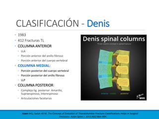
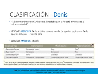
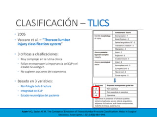
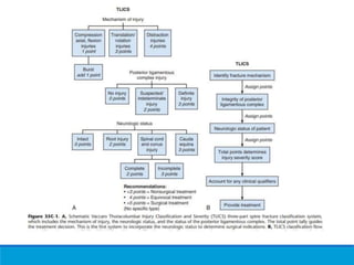
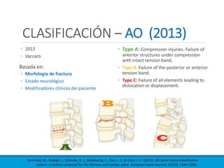
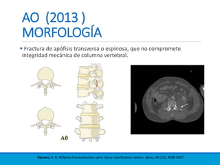
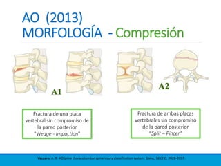
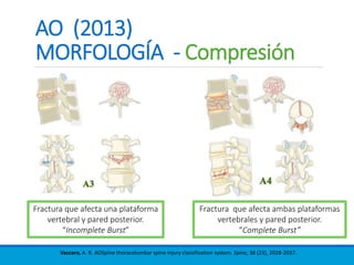
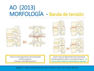
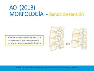
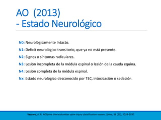
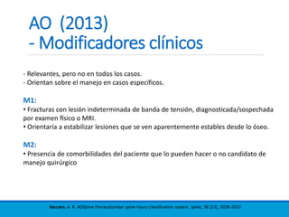
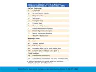
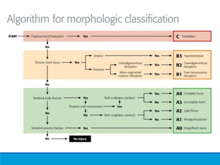
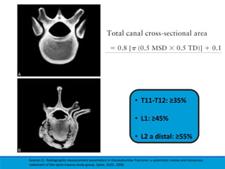
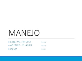
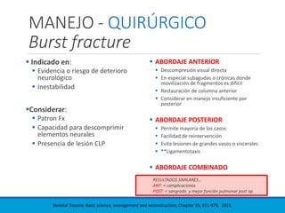
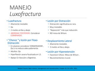
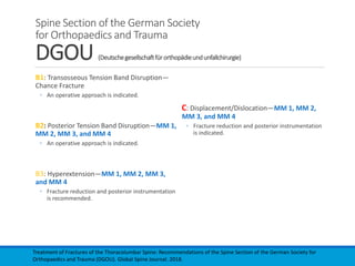
FRACTURAS
TORACOLUMBARES
SEMINARIOS COLUMNA
D R . P E D R O O S O R I O V A L D I V I A
R E S I D E N T E O R T O P E D I A & T R A U M A T O L O G Í A
U N I V E R S I D A D D E L O S A N D E S
2 0 1 9
- CLASIFICACIONES
- ESTUDIO IMAGENOLÓGICO
- MANEJO
FRACTURAS TORACOLUMBARES
CLASIFICACIÓN
• Boehler (1929): Mecanismo y Geometría
• Fractura compresiva
• Compresión del cuerpo y distracción de elementos posteriores
• Lesión en extensión
• Fractura por corte
• Lesión rotacional
• Watson Jones (1938): Incluye el concepto de Estabilidad
• “Integrity of the posterior ligamentous complex (PLC) is essential for stability“
• 4
• Fx en cuña simple – Fx conminuta – Luxofractura – Lesión por hiperextensión
• Sir Frank Holdsworth (1970): Incluye el concepto de 2 Columnas
• Cuerpo vertebral + Disco Y Articulaciones facetarias + PLC
• N: 1000
Azam MQ, Sadat-Ali M. The Concept of Evolution of Thoracolumbar Fracture Classifications Helps in Surgical
Decisions. Asian Spine J. 2015;9(6):984–994.
CLASIFICACIÓN - Denis
◦ 1983
◦ 412 Fracturas TL
◦ COLUMNA ANTERIOR
◦ LLA
◦ Porción anterior del anillo fibroso
◦ Porción anterior del cuerpo vertebral
◦ COLUMNA MEDIAL:
◦ Porción posterior del cuerpo vertebral
◦ Porción posterior del anillo fibroso
◦ LLP
◦ COLUMNA POSTERIOR:
◦ Complejo lig. posterior: Amarillo,
Supraespinoso, Interespinoso
◦ Articulaciones facetarias
Azam MQ, Sadat-Ali M. The Concept of Evolution of Thoracolumbar Fracture Classifications Helps in Surgical
Decisions. Asian Spine J. 2015;9(6):984–994.
CLASIFICACIÓN - Denis
◦ “ Sólo compromiso de CLP no lleva a inestabilidad, si no está involucrada la
columna medial”.
◦ LESIONES MENORES: Fx de apófisis transversa – Fx de apófisis espinosa – Fx de
apófisis articular – Fx de la pars
◦ LESIONES MAYORES: 4 tipos:
Azam MQ, Sadat-Ali M. The Concept of Evolution of Thoracolumbar Fracture Classifications Helps in Surgical
Decisions. Asian Spine J. 2015;9(6):984–994.
CLASIFICACIÓN – AO /Magerl
◦ 1994
◦ N: 1445
◦ Sistema basado en 3 principales
mecanismos de daño:
◦ COMPRESIÓN
◦ DISTRACCIÓN o LESIÓN
BANDA DE TENSIÓN
◦ ROTACIÓN
Azam MQ, Sadat-Ali M. The Concept of Evolution of Thoracolumbar Fracture Classifications Helps in Surgical
Decisions. Asian Spine J. 2015;9(6):984–994.
CLASIFICACIÓN – TLICS
◦ 2005
◦ Vaccaro et al. – “Thoraco-lumbar
injury classification system”
◦ 3 críticas a clasificaciones:
◦ Muy complejas en la rutina clínica
◦ Fallan en reconocer la importancia del CLP y el
estado neurológico
◦ No sugieren opciones de tratamiento
◦ Basado en 3 variables:
◦ Morfología de la Fractura
◦ Integridad del CLP
◦ Estado neurológico del paciente
Azam MQ, Sadat-Ali M. The Concept of Evolution of Thoracolumbar Fracture Classifications Helps in Surgical
Decisions. Asian Spine J. 2015;9(6):984–994.
CLASIFICACIÓN – AO (2013)
◦ 2013
◦ Vaccaro
Basada en:
◦ Morfología de fractura
◦ Estado neurológico
◦ Modificadores clínicos del paciente
◦ Type A: Compression injuries. Failure of
anterior structures under compression
with intact tension band.
◦ Type B: Failure of the posterior or anterior
tension band.
◦ Type C: Failure of all elements leading to
dislocation or displacement.
Reinhold, M., Audigé, L., Schnake, K. J., Bellabarba, C., Dai, L.-Y., & Oner, F. C. (2013). AO spine injury classification
system: a revision proposal for the thoracic and lumbar spine. European Spine Journal, 22(10), 2184–2201.
AO (2013 )
MORFOLOGÍA
 Fractura de apófisis transversa o espinosa, que no compromete
integridad mecánica de columna vertebral.
Vaccaro, A. R. AOSpine thoracolumbar spine injury classification system. Spine, 38 (23), 2028-2037.
AO (2013)
MORFOLOGÍA - Compresión
Vaccaro, A. R. AOSpine thoracolumbar spine injury classification system. Spine, 38 (23), 2028-2037.
Fractura de una placa
vertebral sin compromiso de
la pared posterior
“Wedge - Impaction”
Fractura de ambas placas
vertebrales sin compromiso
de la pared posterior
“Split – Pincer”
AO (2013)
MORFOLOGÍA - Compresión
Vaccaro, A. R. AOSpine thoracolumbar spine injury classification system. Spine, 38 (23), 2028-2037.
Fractura que afecta una plataforma
vertebral y pared posterior.
“Incomplete Burst”
Fractura que afecta ambas plataformas
vertebrales y pared posterior.
“Complete Burst”
AO (2013)
MORFOLOGÍA - Banda de tensión
Vaccaro, A. R. AOSpine thoracolumbar spine injury classification system. Spine, 38 (23), 2028-2037.
Lesión osea y/o ligamentaria de la banda de
tensión posterior junto a una fractura tipo A. La
fractura tipo A debe ser clasificada por separado.
Fractura monosegmentaria de la banda de
tensión posterior
(“Chance o Seat Belt fracture”)
AO (2013)
MORFOLOGÍA - Banda de tensión
Vaccaro, A. R. AOSpine thoracolumbar spine injury classification system. Spine, 38 (23), 2028-2037.
Hiperextensión: Lesión de banda de
tensión anterior por cuerpo o disco
vertebral. Visagra posterior intacta.
AO (2013)
MORFOLOGÍA - Luxofractura
Vaccaro, A. R. AOSpine thoracolumbar spine injury classification system. Spine, 38 (23), 2028-2037.
Fracturas desplazadas, con traslación o
rotación.
Se puede asociar a tipos A y/o B.
AO (2013)
- Estado Neurológico
Vaccaro, A. R. AOSpine thoracolumbar spine injury classification system. Spine, 38 (23), 2028-2037.
N0: Neurológicamente Intacto.
N1: Deficit neurológico transitorio, que ya no está presente.
N2: Signos o síntomas radiculares.
N3: Lesión incompleta de la médula espinal o lesión de la cauda equina.
N4: Lesión completa de la médula espinal.
Nx: Estado neurológico desconocido por TEC, intoxicación o sedación.
AO (2013)
- Modificadores clínicos
Vaccaro, A. R. AOSpine thoracolumbar spine injury classification system. Spine, 38 (23), 2028-2037.
- Relevantes, pero no en todos los casos.
- Orientan sobre el manejo en casos específicos.
M1:
• Fracturas con lesión indeterminada de banda de tensión, diagnosticada/sospechada
por examen físico o MRI.
• Orientaría a estabilizar lesiones que se ven aparentemente estables desde lo óseo.
M2:
• Presencia de comorbilidades del paciente que lo pueden hacer o no candidato de
manejo quirúrgico
Skeletal Trauma: Basic science, management and reconstruction, Chapter 35, 911-979. 2015.
IMAGEN
• FRACTURAS POR ESTALLIDO Y LUXOFRACTURAS
• MEDICIONES
IMAGEN
Burst fracture
• RX o TC AP:
• Aumento de distancia interpedicular
comparado con niveles adyacentes.
• RX Lateral o TC sagital:
• Diminucion altura vertebral
• Falta de definición cortical
• Retropulsión de muro posterior
• Cifosis local
• Aumento distancia interespinosa (Sospecha
lesión CLP)
• TC:
• Patrón de la fractura
• Evaluación inestabilidad (amplitud de art.
Facetarias)
• Compromiso del canal raquideo
Skeletal Trauma: Basic science, management and reconstruction, Chapter 35, 911-979. 2015.
IMAGEN
Burst fracture
• RNM:
Vaccaro: 12 criterios para evaluar disrrupción
del CLP.
• Edema posterior en STIR
• Disrrupción de lig. Amarillo, IE y /o SE
• Cifosis local sin lesión vertebral
• Espacio interespinoso aumentado respecto a
adyacentes en Coronal
• Gap interespinoso palpable
• Sensibilidal posterior focal al examen físico
• Diastasis de art. facetarias
• Fractura avulsiva de la espina superior o
inferior
• Mecanismo lesional
• Compresión >50% de cuerpo vertebral
anterior en Lateral sin Fx de pared posterior en
CT
• Luxación vertebral
• Baja especificidad para lesión CLP
= Falsos positivos
Skeletal Trauma: Basic science, management and reconstruction, Chapter 35, 911-979. 2015.
IMAGEN
Luxofractura
• RX o TC:
• Desplazamiento de vertebra cefálica en
relación a la inferior.
• Articulaciones facetarias incongruentes.
• RNM:
• Determinar lesión nerviosa, en especial en
nivel neurológico comprometido no
relacionado con nivel de Fx.
• Acotar lesion de CLP.
o Elementos posteriores SIEMPRE
comprometidos.
o Fáciles de reconocer.
o SIEMPRE inestables.
Skeletal Trauma: Basic science, management and reconstruction, Chapter 35, 911-979. 2015.
Mediciones
Radiográficas
1. ALINEACIÓN SAGITAL
◦ Ángulo de Cobb (>15-20º)
◦ Deformidad segmentaria de Gardner
◦ Desplazamiento cuerpo vertebral
◦ Índice sagital
2. PORCENTAJE COMPRESIÓN CUERPO VERTEBRAL ANTERIOR
◦ >50% = Asociación a complicaciones en manejo CONSERVADOR
◦
3. DIMENSIÓN CANAL MEDULAR
◦ Área transversal del canal (Mejor correlación con outcome neuro.)
◦ Porcentaje de oclusión del canal: En base a: Áreas transversales o Diámetros sagitales
◦ Relación Sagital a Transverso
Keynan O.: Radiographic measurement parameters in thoracolumbar fractures: a systematic review and consensus
statement of the spine trauma study group. Spine, 31(5). 2006.
Keynan O.: Radiographic measurement parameters in thoracolumbar fractures: a systematic review and consensus
statement of the spine trauma study group. Spine, 31(5). 2006.
Keynan O.: Radiographic measurement parameters in thoracolumbar fractures: a systematic review and consensus
statement of the spine trauma study group. Spine, 31(5). 2006.
• T11-T12: ≥35%
• L1: ≥45%
• L2 a distal: ≥55%
Keynan O.: Radiographic measurement parameters in thoracolumbar fractures: a systematic review and consensus
statement of the spine trauma study group. Spine, 31(5). 2006.
MANEJO
o SKELETAL TRAUMA ( 2 0 1 5 )
o AOSPINE - TL AOSIS ( 2 0 1 5 )
o DGOU ( 2 0 1 8 )
MANEJO - CONSERVADOR
Burst fracture
• Fx estable (sin lesión significativa de
CLP)
• Ausencia de déficit neurológico
• Ortesis espinal personalizada
• Corset Jewett (hiperextensión) (T5-
T10)
• Studies found that bracing had no effect on
pain, radiologic results, kyphotic angle or
functional outcome. ¿?
• Considerar ortesis:
• Fx lumbares bajas
• Fx TL con dolor intratable
• Riesgo de poca adherencia a indicaciones
• Fx torácica baja con riesgo de Cifosis
• Indicar:
• Deambulación precoz
• Evitar actividades de riesgo por 3-6 meses
• Control Urgencia SOS
• Seguimiento Rx: Cifosis>30º o Escoliosis >10º
Shamji MF, Roffey DM, Young DK, et al: A Pilot Evaluation of the Role of Bracing in Stable Thoracolumbar Burst Fractures
Without Neurologic Deficit. J Spinal Disord Tech Aug 18, 2012
MANEJO - QUIRÚRGICO
Burst fracture
 Indicado en:
 Evidencia o riesgo de deterioro
neurológico
 Inestabilidad
Considerar:
 Patron Fx
 Capacidad para descomprimir
elementos neurales
 Presencia de lesión CLP
 ABORDAJE ANTERIOR
 Descompresión visual directa
 En especial subagudas o crónicas donde
movilización de fragmentos es difícil
 Restauración de columna anterior
 Considerar en manejo insuficiente por
posterior
 ABORDAJE POSTERIOR
 Permite mayoría de los casos
 Facilidad de reintervención
 Evita lesiones de grandes vasos o viscerales
 **Ligamentotaxis
 ABORDAJE COMBINADO
RESULTADOS SIMILARES…
ANT: < complicaciones
POST: < sangrado y mejor función pulmonar post op.
Skeletal Trauma: Basic science, management and reconstruction, Chapter 35, 911-979. 2015.
MANEJO
Luxofractura
• Luxofractura
• Altamente inestable
• Qx
• 2 niveles arriba y abajo
• ABORDAJE POSTERIOR. Considerar
COMBINADO.
• “Chance “ y lesión por Flexo-
Distracción
• En jóvenes considerar CONSERVADOR ,
Qx si no reduce adecuadamente.
• Adultos: Qx.
• Hiperflexión severa focalizada en L3.
• Apoyo Cx Vascular y Digestivo.
• Lesión por Distracción
• Distracción significativa es rara.
• Muy inestable.
• “Reposición” más que reducción.
• NO mesa de Wilson.
• Desplazamiento Lateral
• Altamente inestable.
• 3 niveles arriba y abajo.
• Lesión por Hiperextensión
• Favorecer Cifosis: Mesa de Wilson .
• Neuromonitoreo crucial.
Skeletal Trauma: Basic science, management and reconstruction, Chapter 35, 911-979. 2015.
Nonoperative treatment versus Operative
treatment for Thoracolumbar Burst fractures
without neurologic deficit
 Follow-up time: 24 to 118 months.
 No differences in pain.
 At approximately 4 years, the operative group had an improvement in kyphosis
from baseline by 1.8 degrees with a mean of 11 degrees, as opposed to a
kyphosis of 16 degrees with a worsening of 3.3 degrees in the nonoperative
group.
 There was also no association found between kyphosis and pain.
 Operative treatment was also associated with higher complication rates and
costs.
 Return to work rate were statistically insignificant difference.
Gnanenthiran SR, Adie S, Harris IA: Nonoperative versus operative treatment for thoracolumbar burst fractures without
neurologic deficit: a meta-analysis. Clin Orthop Relat Res 470(2):567–577, 2012
Comparison of Posterior Instrumentation
with and without Fusion in Burst fracture
 Meta-analysis of four studies including 220 patients.
 NO difference at follow-up was present between groups.
 Operating time and intraoperative blood loss were less in the fusion-less
group.
 No differences were present in rates of hardware failure or surgical
complications.
Tian NF, Wu YS, Zhang XL, et al: Fusion versus nonfusion for surgically treated thoracolumbar burst fractures: a meta-
analysis. PLoS One 8(5). 2013.
Thoracolumbar AOSpine Injury System
TL AOSIS
3 o menos = CONSERVADOR
5 o más = QUIRÚRGICO
Vaccaro A.: The surgical algorithm for the AOSpine thoracolumbar spine injury classification system.
Eur Spine Journal 25(4). 2015.
Thoracolumbar AOSpine Injury System
TL AOSIS
 Algoritmo elaborado en base a
respuestas de miembros de
AOSpine, frente a Fx TL
controversiales.
 N: 483
 2 a 8 puntos.
 Variabilidad regional en 15 de 19
casos.
¿Indicación Qx?
 30% o menos = CONSERVADOR
 70% o más = QUIRÚRGICO
Vaccaro A.: The surgical algorithm for the AOSpine thoracolumbar spine injury classification system.
Eur Spine Journal 25(4). 2015.
Spine Section of the German Society
for Orthopaedics and Trauma
DGOU (Deutschegesellschaftfürorthopädieundunfallchirurgie)
MORFOLOGICAL MODIFIERS
•MM1 = Sagittal and coronal alignment of spine
• EPA (Endplate angle): and Sagital angle:
• δEPA: EPA + cifosis lumbar o - lordosis lumbar.
• <15-20º = CONSERVADOR
• >15-20º = QUIRÚRGICO
• MM2 = Comminution of vertebral body
• McCormack (1976): 1/3 de craneal a caudal.
• MM3 = Stenosis of spinal canal
• % perdido en relación a niveles sanos adyacentes
• MM4 = Intervertebral disc lesion
Treatment of Fractures of the Thoracolumbar Spine: Recommendations of the Spine Section of the German Society for
Orthopaedics and Trauma (DGOU). Global Spine Journal. 2018.
Spine Section of the German Society
for Orthopaedics and Trauma
DGOU (Deutschegesellschaftfürorthopädieundunfallchirurgie)
A1: Wedge-Compression—MM 1
- δEPA < 5° to 20° conservative therapy.
- δEPA >15° to 20° operative treatment.
A2: Split—MM 2 and MM 4
- Early mobilization, adequate analgesia, and
physiotherapy.
*** Wide fragment separation and/or relevant
lesion of the intervertebral disc can be an
indication for surgery.
A3: Incomplete Burst—MM 1, MM 2, MM 3, and
MM 4
- δEPA <15° to 20° and/or scoliosis <10°
conservative therapy.
- δEPA >15° to 20° and/or scoliosis >10° operative
treatment.
A4: Complete Burst—MM 1, MM 2, MM 3, and
MM 4
- δEPA <15° to 20° and/or scoliosis <10°
conservative therapy.
- δEPA > 15° to 20° and/or scoliosis >10° operative
treatment.
- It is possible that there is gross fragment
displacement with critical narrowing of the spinal
canal without considerable deviation of δEPA and
scoliosis angle.
Treatment of Fractures of the Thoracolumbar Spine: Recommendations of the Spine Section of the German Society for
Orthopaedics and Trauma (DGOU). Global Spine Journal. 2018.
Spine Section of the German Society
for Orthopaedics and Trauma
DGOU (Deutschegesellschaftfürorthopädieundunfallchirurgie)
B1: Transosseous Tension Band Disruption—
Chance Fracture
◦ An operative approach is indicated.
B2: Posterior Tension Band Disruption—MM 1,
MM 2, MM 3, and MM 4
◦ An operative approach is indicated.
B3: Hyperextension—MM 1, MM 2, MM 3,
and MM 4
◦ Fracture reduction and posterior instrumentation
is recommended.
C: Displacement/Dislocation—MM 1, MM 2,
MM 3, and MM 4
◦ Fracture reduction and posterior instrumentation
is indicated.
Treatment of Fractures of the Thoracolumbar Spine: Recommendations of the Spine Section of the German Society for
Orthopaedics and Trauma (DGOU). Global Spine Journal. 2018.
Take home messages…
1. La mejor clasificación es la que orienta al manejo, es reproducible y
tiene la menor variabilidad interobservador.
2. La medición radiológica debe ser estandarizada.
3. Para decidir el manejo adecuado, siempre hay que considerar los
modificadores clínicos y morfológicos.
BIBLIOGRAFÍA
1. Azam MQ, Sadat-Ali M. The Concept of Evolution of Thoracolumbar Fracture Classifications Helps in
Surgical Decisions. Asian Spine J. 9(6):984–994. 2015.
2. Reinhold, M., Audigé, L., Schnake, K. J., Bellabarba, C., Dai, L.-Y., & Oner, F. C. (2013). AO spine injury
classification system: a revision proposal for the thoracic and lumbar spine. Eur Spine Journal,
22(10), 2184–2201.
3. Vaccaro, A. R. AOSpine thoracolumbar spine injury classification system. Spine, 38 (23). 2013.
4. Campbell's Operative Orthopaedics, Chapter 41, 1801-1811. 2017.
5. Skeletal Trauma: Basic science, management and reconstruction, Chapter 35, 911-979. 2015.
6. Vaccaro, A. R.: The surgical algorithm for the AOSpine thoracolumbar spine injury classification
system. Eur Spine Journal ,25(4). 2015.
7. Akhil P. Verheyden: Treatment of Fractures of the Thoracolumbar Spine: Recommendations of the
Spine Section of the German Society for Orthopaedics and Trauma (DGOU). Global Spine Journal,
8(2). 2018.
8. Keynan O.: Radiographic measurement parameters in thoracolumbar fractures: a systematic review
and consensus statement of the spine trauma study group. Spine, 31(5). 2006.
FRACTURAS
TORACOLUMBARES
SEMINARIOS COLUMNA
D R . P E D R O O S O R I O V A L D I V I A
R E S I D E N T E O R T O P E D I A & T R A U M A T O L O G Í A
U N I V E R S I D A D D E L O S A N D E S
2 0 1 9

Fracturas Toracolumbares

  • 1.
    FRACTURAS TORACOLUMBARES SEMINARIOS COLUMNA D R. P E D R O O S O R I O V A L D I V I A R E S I D E N T E O R T O P E D I A & T R A U M A T O L O G Í A U N I V E R S I D A D D E L O S A N D E S 2 0 1 9
  • 2.
    - CLASIFICACIONES - ESTUDIOIMAGENOLÓGICO - MANEJO FRACTURAS TORACOLUMBARES
  • 3.
    CLASIFICACIÓN • Boehler (1929):Mecanismo y Geometría • Fractura compresiva • Compresión del cuerpo y distracción de elementos posteriores • Lesión en extensión • Fractura por corte • Lesión rotacional • Watson Jones (1938): Incluye el concepto de Estabilidad • “Integrity of the posterior ligamentous complex (PLC) is essential for stability“ • 4 • Fx en cuña simple – Fx conminuta – Luxofractura – Lesión por hiperextensión • Sir Frank Holdsworth (1970): Incluye el concepto de 2 Columnas • Cuerpo vertebral + Disco Y Articulaciones facetarias + PLC • N: 1000 Azam MQ, Sadat-Ali M. The Concept of Evolution of Thoracolumbar Fracture Classifications Helps in Surgical Decisions. Asian Spine J. 2015;9(6):984–994.
  • 4.
    CLASIFICACIÓN - Denis ◦1983 ◦ 412 Fracturas TL ◦ COLUMNA ANTERIOR ◦ LLA ◦ Porción anterior del anillo fibroso ◦ Porción anterior del cuerpo vertebral ◦ COLUMNA MEDIAL: ◦ Porción posterior del cuerpo vertebral ◦ Porción posterior del anillo fibroso ◦ LLP ◦ COLUMNA POSTERIOR: ◦ Complejo lig. posterior: Amarillo, Supraespinoso, Interespinoso ◦ Articulaciones facetarias Azam MQ, Sadat-Ali M. The Concept of Evolution of Thoracolumbar Fracture Classifications Helps in Surgical Decisions. Asian Spine J. 2015;9(6):984–994.
  • 5.
    CLASIFICACIÓN - Denis ◦“ Sólo compromiso de CLP no lleva a inestabilidad, si no está involucrada la columna medial”. ◦ LESIONES MENORES: Fx de apófisis transversa – Fx de apófisis espinosa – Fx de apófisis articular – Fx de la pars ◦ LESIONES MAYORES: 4 tipos: Azam MQ, Sadat-Ali M. The Concept of Evolution of Thoracolumbar Fracture Classifications Helps in Surgical Decisions. Asian Spine J. 2015;9(6):984–994.
  • 6.
    CLASIFICACIÓN – AO/Magerl ◦ 1994 ◦ N: 1445 ◦ Sistema basado en 3 principales mecanismos de daño: ◦ COMPRESIÓN ◦ DISTRACCIÓN o LESIÓN BANDA DE TENSIÓN ◦ ROTACIÓN Azam MQ, Sadat-Ali M. The Concept of Evolution of Thoracolumbar Fracture Classifications Helps in Surgical Decisions. Asian Spine J. 2015;9(6):984–994.
  • 7.
    CLASIFICACIÓN – TLICS ◦2005 ◦ Vaccaro et al. – “Thoraco-lumbar injury classification system” ◦ 3 críticas a clasificaciones: ◦ Muy complejas en la rutina clínica ◦ Fallan en reconocer la importancia del CLP y el estado neurológico ◦ No sugieren opciones de tratamiento ◦ Basado en 3 variables: ◦ Morfología de la Fractura ◦ Integridad del CLP ◦ Estado neurológico del paciente Azam MQ, Sadat-Ali M. The Concept of Evolution of Thoracolumbar Fracture Classifications Helps in Surgical Decisions. Asian Spine J. 2015;9(6):984–994.
  • 9.
    CLASIFICACIÓN – AO(2013) ◦ 2013 ◦ Vaccaro Basada en: ◦ Morfología de fractura ◦ Estado neurológico ◦ Modificadores clínicos del paciente ◦ Type A: Compression injuries. Failure of anterior structures under compression with intact tension band. ◦ Type B: Failure of the posterior or anterior tension band. ◦ Type C: Failure of all elements leading to dislocation or displacement. Reinhold, M., Audigé, L., Schnake, K. J., Bellabarba, C., Dai, L.-Y., & Oner, F. C. (2013). AO spine injury classification system: a revision proposal for the thoracic and lumbar spine. European Spine Journal, 22(10), 2184–2201.
  • 10.
    AO (2013 ) MORFOLOGÍA Fractura de apófisis transversa o espinosa, que no compromete integridad mecánica de columna vertebral. Vaccaro, A. R. AOSpine thoracolumbar spine injury classification system. Spine, 38 (23), 2028-2037.
  • 11.
    AO (2013) MORFOLOGÍA -Compresión Vaccaro, A. R. AOSpine thoracolumbar spine injury classification system. Spine, 38 (23), 2028-2037. Fractura de una placa vertebral sin compromiso de la pared posterior “Wedge - Impaction” Fractura de ambas placas vertebrales sin compromiso de la pared posterior “Split – Pincer”
  • 12.
    AO (2013) MORFOLOGÍA -Compresión Vaccaro, A. R. AOSpine thoracolumbar spine injury classification system. Spine, 38 (23), 2028-2037. Fractura que afecta una plataforma vertebral y pared posterior. “Incomplete Burst” Fractura que afecta ambas plataformas vertebrales y pared posterior. “Complete Burst”
  • 13.
    AO (2013) MORFOLOGÍA -Banda de tensión Vaccaro, A. R. AOSpine thoracolumbar spine injury classification system. Spine, 38 (23), 2028-2037. Lesión osea y/o ligamentaria de la banda de tensión posterior junto a una fractura tipo A. La fractura tipo A debe ser clasificada por separado. Fractura monosegmentaria de la banda de tensión posterior (“Chance o Seat Belt fracture”)
  • 14.
    AO (2013) MORFOLOGÍA -Banda de tensión Vaccaro, A. R. AOSpine thoracolumbar spine injury classification system. Spine, 38 (23), 2028-2037. Hiperextensión: Lesión de banda de tensión anterior por cuerpo o disco vertebral. Visagra posterior intacta.
  • 15.
    AO (2013) MORFOLOGÍA -Luxofractura Vaccaro, A. R. AOSpine thoracolumbar spine injury classification system. Spine, 38 (23), 2028-2037. Fracturas desplazadas, con traslación o rotación. Se puede asociar a tipos A y/o B.
  • 16.
    AO (2013) - EstadoNeurológico Vaccaro, A. R. AOSpine thoracolumbar spine injury classification system. Spine, 38 (23), 2028-2037. N0: Neurológicamente Intacto. N1: Deficit neurológico transitorio, que ya no está presente. N2: Signos o síntomas radiculares. N3: Lesión incompleta de la médula espinal o lesión de la cauda equina. N4: Lesión completa de la médula espinal. Nx: Estado neurológico desconocido por TEC, intoxicación o sedación.
  • 17.
    AO (2013) - Modificadoresclínicos Vaccaro, A. R. AOSpine thoracolumbar spine injury classification system. Spine, 38 (23), 2028-2037. - Relevantes, pero no en todos los casos. - Orientan sobre el manejo en casos específicos. M1: • Fracturas con lesión indeterminada de banda de tensión, diagnosticada/sospechada por examen físico o MRI. • Orientaría a estabilizar lesiones que se ven aparentemente estables desde lo óseo. M2: • Presencia de comorbilidades del paciente que lo pueden hacer o no candidato de manejo quirúrgico
  • 18.
    Skeletal Trauma: Basicscience, management and reconstruction, Chapter 35, 911-979. 2015.
  • 20.
    IMAGEN • FRACTURAS PORESTALLIDO Y LUXOFRACTURAS • MEDICIONES
  • 21.
    IMAGEN Burst fracture • RXo TC AP: • Aumento de distancia interpedicular comparado con niveles adyacentes. • RX Lateral o TC sagital: • Diminucion altura vertebral • Falta de definición cortical • Retropulsión de muro posterior • Cifosis local • Aumento distancia interespinosa (Sospecha lesión CLP) • TC: • Patrón de la fractura • Evaluación inestabilidad (amplitud de art. Facetarias) • Compromiso del canal raquideo Skeletal Trauma: Basic science, management and reconstruction, Chapter 35, 911-979. 2015.
  • 22.
    IMAGEN Burst fracture • RNM: Vaccaro:12 criterios para evaluar disrrupción del CLP. • Edema posterior en STIR • Disrrupción de lig. Amarillo, IE y /o SE • Cifosis local sin lesión vertebral • Espacio interespinoso aumentado respecto a adyacentes en Coronal • Gap interespinoso palpable • Sensibilidal posterior focal al examen físico • Diastasis de art. facetarias • Fractura avulsiva de la espina superior o inferior • Mecanismo lesional • Compresión >50% de cuerpo vertebral anterior en Lateral sin Fx de pared posterior en CT • Luxación vertebral • Baja especificidad para lesión CLP = Falsos positivos Skeletal Trauma: Basic science, management and reconstruction, Chapter 35, 911-979. 2015.
  • 23.
    IMAGEN Luxofractura • RX oTC: • Desplazamiento de vertebra cefálica en relación a la inferior. • Articulaciones facetarias incongruentes. • RNM: • Determinar lesión nerviosa, en especial en nivel neurológico comprometido no relacionado con nivel de Fx. • Acotar lesion de CLP. o Elementos posteriores SIEMPRE comprometidos. o Fáciles de reconocer. o SIEMPRE inestables. Skeletal Trauma: Basic science, management and reconstruction, Chapter 35, 911-979. 2015.
  • 24.
    Mediciones Radiográficas 1. ALINEACIÓN SAGITAL ◦Ángulo de Cobb (>15-20º) ◦ Deformidad segmentaria de Gardner ◦ Desplazamiento cuerpo vertebral ◦ Índice sagital 2. PORCENTAJE COMPRESIÓN CUERPO VERTEBRAL ANTERIOR ◦ >50% = Asociación a complicaciones en manejo CONSERVADOR ◦ 3. DIMENSIÓN CANAL MEDULAR ◦ Área transversal del canal (Mejor correlación con outcome neuro.) ◦ Porcentaje de oclusión del canal: En base a: Áreas transversales o Diámetros sagitales ◦ Relación Sagital a Transverso Keynan O.: Radiographic measurement parameters in thoracolumbar fractures: a systematic review and consensus statement of the spine trauma study group. Spine, 31(5). 2006.
  • 25.
    Keynan O.: Radiographicmeasurement parameters in thoracolumbar fractures: a systematic review and consensus statement of the spine trauma study group. Spine, 31(5). 2006.
  • 26.
    Keynan O.: Radiographicmeasurement parameters in thoracolumbar fractures: a systematic review and consensus statement of the spine trauma study group. Spine, 31(5). 2006.
  • 27.
    • T11-T12: ≥35% •L1: ≥45% • L2 a distal: ≥55% Keynan O.: Radiographic measurement parameters in thoracolumbar fractures: a systematic review and consensus statement of the spine trauma study group. Spine, 31(5). 2006.
  • 28.
    MANEJO o SKELETAL TRAUMA( 2 0 1 5 ) o AOSPINE - TL AOSIS ( 2 0 1 5 ) o DGOU ( 2 0 1 8 )
  • 29.
    MANEJO - CONSERVADOR Burstfracture • Fx estable (sin lesión significativa de CLP) • Ausencia de déficit neurológico • Ortesis espinal personalizada • Corset Jewett (hiperextensión) (T5- T10) • Studies found that bracing had no effect on pain, radiologic results, kyphotic angle or functional outcome. ¿? • Considerar ortesis: • Fx lumbares bajas • Fx TL con dolor intratable • Riesgo de poca adherencia a indicaciones • Fx torácica baja con riesgo de Cifosis • Indicar: • Deambulación precoz • Evitar actividades de riesgo por 3-6 meses • Control Urgencia SOS • Seguimiento Rx: Cifosis>30º o Escoliosis >10º Shamji MF, Roffey DM, Young DK, et al: A Pilot Evaluation of the Role of Bracing in Stable Thoracolumbar Burst Fractures Without Neurologic Deficit. J Spinal Disord Tech Aug 18, 2012
  • 30.
    MANEJO - QUIRÚRGICO Burstfracture  Indicado en:  Evidencia o riesgo de deterioro neurológico  Inestabilidad Considerar:  Patron Fx  Capacidad para descomprimir elementos neurales  Presencia de lesión CLP  ABORDAJE ANTERIOR  Descompresión visual directa  En especial subagudas o crónicas donde movilización de fragmentos es difícil  Restauración de columna anterior  Considerar en manejo insuficiente por posterior  ABORDAJE POSTERIOR  Permite mayoría de los casos  Facilidad de reintervención  Evita lesiones de grandes vasos o viscerales  **Ligamentotaxis  ABORDAJE COMBINADO RESULTADOS SIMILARES… ANT: < complicaciones POST: < sangrado y mejor función pulmonar post op. Skeletal Trauma: Basic science, management and reconstruction, Chapter 35, 911-979. 2015.
  • 31.
    MANEJO Luxofractura • Luxofractura • Altamenteinestable • Qx • 2 niveles arriba y abajo • ABORDAJE POSTERIOR. Considerar COMBINADO. • “Chance “ y lesión por Flexo- Distracción • En jóvenes considerar CONSERVADOR , Qx si no reduce adecuadamente. • Adultos: Qx. • Hiperflexión severa focalizada en L3. • Apoyo Cx Vascular y Digestivo. • Lesión por Distracción • Distracción significativa es rara. • Muy inestable. • “Reposición” más que reducción. • NO mesa de Wilson. • Desplazamiento Lateral • Altamente inestable. • 3 niveles arriba y abajo. • Lesión por Hiperextensión • Favorecer Cifosis: Mesa de Wilson . • Neuromonitoreo crucial. Skeletal Trauma: Basic science, management and reconstruction, Chapter 35, 911-979. 2015.
  • 32.
    Nonoperative treatment versusOperative treatment for Thoracolumbar Burst fractures without neurologic deficit  Follow-up time: 24 to 118 months.  No differences in pain.  At approximately 4 years, the operative group had an improvement in kyphosis from baseline by 1.8 degrees with a mean of 11 degrees, as opposed to a kyphosis of 16 degrees with a worsening of 3.3 degrees in the nonoperative group.  There was also no association found between kyphosis and pain.  Operative treatment was also associated with higher complication rates and costs.  Return to work rate were statistically insignificant difference. Gnanenthiran SR, Adie S, Harris IA: Nonoperative versus operative treatment for thoracolumbar burst fractures without neurologic deficit: a meta-analysis. Clin Orthop Relat Res 470(2):567–577, 2012
  • 33.
    Comparison of PosteriorInstrumentation with and without Fusion in Burst fracture  Meta-analysis of four studies including 220 patients.  NO difference at follow-up was present between groups.  Operating time and intraoperative blood loss were less in the fusion-less group.  No differences were present in rates of hardware failure or surgical complications. Tian NF, Wu YS, Zhang XL, et al: Fusion versus nonfusion for surgically treated thoracolumbar burst fractures: a meta- analysis. PLoS One 8(5). 2013.
  • 34.
    Thoracolumbar AOSpine InjurySystem TL AOSIS 3 o menos = CONSERVADOR 5 o más = QUIRÚRGICO Vaccaro A.: The surgical algorithm for the AOSpine thoracolumbar spine injury classification system. Eur Spine Journal 25(4). 2015.
  • 35.
    Thoracolumbar AOSpine InjurySystem TL AOSIS  Algoritmo elaborado en base a respuestas de miembros de AOSpine, frente a Fx TL controversiales.  N: 483  2 a 8 puntos.  Variabilidad regional en 15 de 19 casos. ¿Indicación Qx?  30% o menos = CONSERVADOR  70% o más = QUIRÚRGICO Vaccaro A.: The surgical algorithm for the AOSpine thoracolumbar spine injury classification system. Eur Spine Journal 25(4). 2015.
  • 36.
    Spine Section ofthe German Society for Orthopaedics and Trauma DGOU (Deutschegesellschaftfürorthopädieundunfallchirurgie) MORFOLOGICAL MODIFIERS •MM1 = Sagittal and coronal alignment of spine • EPA (Endplate angle): and Sagital angle: • δEPA: EPA + cifosis lumbar o - lordosis lumbar. • <15-20º = CONSERVADOR • >15-20º = QUIRÚRGICO • MM2 = Comminution of vertebral body • McCormack (1976): 1/3 de craneal a caudal. • MM3 = Stenosis of spinal canal • % perdido en relación a niveles sanos adyacentes • MM4 = Intervertebral disc lesion Treatment of Fractures of the Thoracolumbar Spine: Recommendations of the Spine Section of the German Society for Orthopaedics and Trauma (DGOU). Global Spine Journal. 2018.
  • 37.
    Spine Section ofthe German Society for Orthopaedics and Trauma DGOU (Deutschegesellschaftfürorthopädieundunfallchirurgie) A1: Wedge-Compression—MM 1 - δEPA < 5° to 20° conservative therapy. - δEPA >15° to 20° operative treatment. A2: Split—MM 2 and MM 4 - Early mobilization, adequate analgesia, and physiotherapy. *** Wide fragment separation and/or relevant lesion of the intervertebral disc can be an indication for surgery. A3: Incomplete Burst—MM 1, MM 2, MM 3, and MM 4 - δEPA <15° to 20° and/or scoliosis <10° conservative therapy. - δEPA >15° to 20° and/or scoliosis >10° operative treatment. A4: Complete Burst—MM 1, MM 2, MM 3, and MM 4 - δEPA <15° to 20° and/or scoliosis <10° conservative therapy. - δEPA > 15° to 20° and/or scoliosis >10° operative treatment. - It is possible that there is gross fragment displacement with critical narrowing of the spinal canal without considerable deviation of δEPA and scoliosis angle. Treatment of Fractures of the Thoracolumbar Spine: Recommendations of the Spine Section of the German Society for Orthopaedics and Trauma (DGOU). Global Spine Journal. 2018.
  • 38.
    Spine Section ofthe German Society for Orthopaedics and Trauma DGOU (Deutschegesellschaftfürorthopädieundunfallchirurgie) B1: Transosseous Tension Band Disruption— Chance Fracture ◦ An operative approach is indicated. B2: Posterior Tension Band Disruption—MM 1, MM 2, MM 3, and MM 4 ◦ An operative approach is indicated. B3: Hyperextension—MM 1, MM 2, MM 3, and MM 4 ◦ Fracture reduction and posterior instrumentation is recommended. C: Displacement/Dislocation—MM 1, MM 2, MM 3, and MM 4 ◦ Fracture reduction and posterior instrumentation is indicated. Treatment of Fractures of the Thoracolumbar Spine: Recommendations of the Spine Section of the German Society for Orthopaedics and Trauma (DGOU). Global Spine Journal. 2018.
  • 39.
    Take home messages… 1.La mejor clasificación es la que orienta al manejo, es reproducible y tiene la menor variabilidad interobservador. 2. La medición radiológica debe ser estandarizada. 3. Para decidir el manejo adecuado, siempre hay que considerar los modificadores clínicos y morfológicos.
  • 40.
    BIBLIOGRAFÍA 1. Azam MQ,Sadat-Ali M. The Concept of Evolution of Thoracolumbar Fracture Classifications Helps in Surgical Decisions. Asian Spine J. 9(6):984–994. 2015. 2. Reinhold, M., Audigé, L., Schnake, K. J., Bellabarba, C., Dai, L.-Y., & Oner, F. C. (2013). AO spine injury classification system: a revision proposal for the thoracic and lumbar spine. Eur Spine Journal, 22(10), 2184–2201. 3. Vaccaro, A. R. AOSpine thoracolumbar spine injury classification system. Spine, 38 (23). 2013. 4. Campbell's Operative Orthopaedics, Chapter 41, 1801-1811. 2017. 5. Skeletal Trauma: Basic science, management and reconstruction, Chapter 35, 911-979. 2015. 6. Vaccaro, A. R.: The surgical algorithm for the AOSpine thoracolumbar spine injury classification system. Eur Spine Journal ,25(4). 2015. 7. Akhil P. Verheyden: Treatment of Fractures of the Thoracolumbar Spine: Recommendations of the Spine Section of the German Society for Orthopaedics and Trauma (DGOU). Global Spine Journal, 8(2). 2018. 8. Keynan O.: Radiographic measurement parameters in thoracolumbar fractures: a systematic review and consensus statement of the spine trauma study group. Spine, 31(5). 2006.
  • 41.
    FRACTURAS TORACOLUMBARES SEMINARIOS COLUMNA D R. P E D R O O S O R I O V A L D I V I A R E S I D E N T E O R T O P E D I A & T R A U M A T O L O G Í A U N I V E R S I D A D D E L O S A N D E S 2 0 1 9

Notas del editor

  • #10 —Type A: Compression injuries. Failure of anterior structures under compression with intact tension band. Type B: Failure of the posterior or anterior tension band. Type C: Failure of all elements leading to dislocation or displacement.
  • #12 CUÑA – IMPACTO // DIVISIÓN - PINZA
  • #13 ESTALLIDO
  • #14 CHANCE OR SEAT BELT FRACTURE
  • #25 Area transversal del canal: Mejor correlación con outcome neurológico % oclusión del canal: en proximas diapos
  • #30 Shamii: estudio con N 23 / T12-L2 / corset ToracoLumbarSacralOrtesis = TLSO / porfavaaar cuando sirve? Estudios son contradictorios
  • #34 Fusión con injerto óseo (fleiderman)