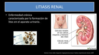
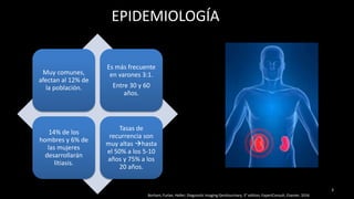
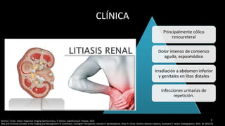
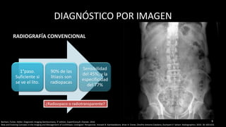
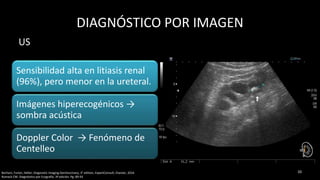
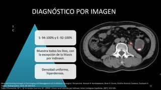
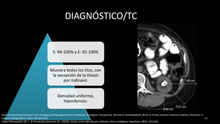
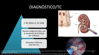
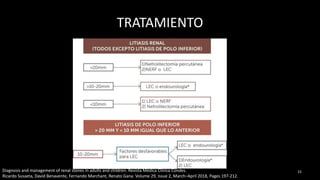
PROFESOR TITULAR
DR. JESUS RAMIREZ MARTINEZ
COORDINADORES DEL MÓDULO:
DR. ALVARO VARGAS C.
DRA. VANESSA ZAMORA P.
DR. RAÚL GÓMEZ B.
DRA. AMEYALI PÉREZ HUITRÓN
RESIDENTE DE SEGUNDO AÑO
IMAGENOLOGÍA DIAGNÓSTICA Y TERAPEÚTICA
1
LITIASIS RENAL
Borhani, Furlan, Heller; Diagnostic Imaging Genitourinary, 3° edition, ExpertConsult, Elsevier, 2016
• Enfermedad crónica
caracterizada por la formación de
litos en el aparato urinario.
2
EPIDEMIOLOGÍA
Muy comunes,
afectan al 12% de
la población.
Es más frecuente
en varones 3:1.
Entre 30 y 60
años.
14% de los
hombres y 6% de
las mujeres
desarrollarán
litiasis.
Tasas de
recurrencia son
muy altas hasta
el 50% a los 5-10
años y 75% a los
20 años.
3
Borhani, Furlan, Heller; Diagnostic Imaging Genitourinary, 3° edition, ExpertConsult, Elsevier, 2016
ETIOLOGÍA
GenéticaDieta
Antecedentes
de infecciones
de repetición
HipercalciuriaGotaHiperoxaluriaCistinuria
4Borhani, Furlan, Heller; Diagnostic Imaging Genitourinary, 3° edition, ExpertConsult, Elsevier, 2016
New and Evolving Concepts in the Imaging and Management of Urolithiasis: Urologists’ Perspective. Avinash R. Kambadakone, Brian H. Eisner, Onofrio Antonio Catalano, Dushyant V. Sahani. Radiographics. 2010. 30: 603-623.
FISIOPATOLOGÍA
La estasis urinaria favorece su formación en casos de
obstrucción crónica
La mayoría compuestos por calcio: mezcla de
oxalato y fosfato cálcico (34%), oxalato de calcio
puro (33%) y fosfato cálcico puro (6%).
“Litos por infección” son causados por gérmenes
gram- (Proteus) productores de ureasa en una
orina alcalina.
55Borhani, Furlan, Heller; Diagnostic Imaging Genitourinary, 3° edition, ExpertConsult, Elsevier, 2016
New and Evolving Concepts in the Imaging and Management of Urolithiasis: Urologists’ Perspective. Avinash R. Kambadakone, Brian H. Eisner, Onofrio Antonio Catalano, Dushyant V. Sahani. Radiographics. 2010. 30: 603-623.
Figure 1 Chart illustrates commonly occurring urinary tract stones and describes their salient features. KUB = kidney, ureter, bladder.
TIPOS DE LITIASIS RENAL
Figure 1 Chart illustrates commonly occurring urinary tract stones and describes their salient features. KUB = kidney, ureter, bladder.
New and Evolving Concepts in the Imaging and Management of Urolithiasis: Urologists’ Perspective. Avinash R. Kambadakone, Brian H. Eisner, Onofrio Antonio Catalano, Dushyant V.
Sahani. Radiographics. 2010. 30: 603-623.
CLÍNICA
Principalmente cólico
renoureteral
Dolor intenso de comienzo
agudo, espasmódico
Irradiación a abdomen inferior
y genitales en litos distales
Infecciones urinarias de
repetición.
77Borhani, Furlan, Heller; Diagnostic Imaging Genitourinary, 3° edition, ExpertConsult, Elsevier, 2016
New and Evolving Concepts in the Imaging and Management of Urolithiasis: Urologists’ Perspective. Avinash R. Kambadakone, Brian H. Eisner, Onofrio Antonio Catalano, Dushyant V. Sahani. Radiographics. 2010. 30: 603-623.
ELIMINACIÓN ESPONTÁNEA
Eliminación espontánea
<4mm 80%
4-6mm 50%
>8mm 20%
8New and Evolving Concepts in the Imaging and Management of Urolithiasis: Urologists’ Perspective. Avinash R. Kambadakone, Brian H. Eisner, Onofrio Antonio Catalano, Dushyant V. Sahani.
Radiographics. 2010. 30: 603-623.
DIAGNÓSTICO POR IMAGEN
RADIOGRAFÍA CONVENCIONAL
1°paso.
Suficiente si
se ve el lito.
90% de las
litiasis son
radiopacas
Sensibilidad
del 45%, y la
especificidad
del 77%
9
¿Radioopaco o radiotransparente?
9Borhani, Furlan, Heller; Diagnostic Imaging Genitourinary, 3° edition, ExpertConsult, Elsevier, 2016
New and Evolving Concepts in the Imaging and Management of Urolithiasis: Urologists’ Perspective. Avinash R. Kambadakone, Brian H. Eisner, Onofrio Antonio Catalano, Dushyant V. Sahani. Radiographics. 2010. 30: 603-623.
DIAGNÓSTICO POR IMAGEN
Sensibilidad alta en litiasis renal
(96%), pero menor en la ureteral.
Imágenes hiperecogénicos →
sombra acústica
Doppler Color → Fenómeno de
Centelleo
1010Borhani, Furlan, Heller; Diagnostic Imaging Genitourinary, 3° edition, ExpertConsult, Elsevier, 2016
Rumack CM. Diagnóstico por Ecografía. 3ª edición. Pg. 89-93
US
DIAGNÓSTICO POR IMAGEN
Litos
(ureterales)
Nefromegalia
Fase nefrográfica
asimétrica, tardía
y persistente.
Hidronefrosis
11
Davidson. Radiología del riñón. Capítulo 17, Dilatación del sistema pielocalicial y el uréter.Pag. 466-468.
UROGRAFÍA EXCRETORA
DIAGNÓSTICO POR IMAGEN
S: 94-100% y E: 92-100%
Muestra todos los litos, con
la excepción de la litiasis
por indinavir.
Densidad uniforme,
hiperdensos.
12
New and Evolving Concepts in the Imaging and Management of Urolithiasis: Urologists’ Perspective. Avinash R. Kambadakone, Brian H. Eisner, Onofrio Antonio Catalano, Dushyant V.
Sahani. Radiographics. 2010. 30: 603-623.
Traba Villameytide, Mª L., & Fernández-Guerrero, M.. (2004). Litiasis renal inducida por Indinavir. Actas Urológicas Españolas, 28(7), 523-526.
T
C
DIAGNÓSTICO/TC
S: 94-100% y E: 92-100%
Muestra todos los litos, con
la excepción de la litiasis
por indinavir.
Densidad uniforme,
hiperdensos.
13
New and Evolving Concepts in the Imaging and Management of Urolithiasis: Urologists’ Perspective. Avinash R. Kambadakone, Brian H. Eisner, Onofrio Antonio Catalano, Dushyant V.
Sahani. Radiographics. 2010. 30: 603-623.
Traba Villameytide, Mª L., & Fernández-Guerrero, M.. (2004). Litiasis renal inducida por Indinavir. Actas Urológicas Españolas, 28(7), 523-526.
DIAGNÓSTICO/TC
S: 94-100% y E: 92-100%
Muestra todos los litos, con
la excepción de la litiasis
por indinavir.
Densidad uniforme,
hiperdensos.
14
New and Evolving Concepts in the Imaging and Management of Urolithiasis: Urologists’ Perspective. Avinash R. Kambadakone, Brian H. Eisner, Onofrio Antonio Catalano, Dushyant V. Sahani.
Radiographics. 2010. 30: 603-623.
Traba Villameytide, Mª L., & Fernández-Guerrero, M.. (2014). Litiasis renal inducida por Indinavir. Actas Urológicas Españolas, 28(7), 523-526.
TRATAMIENTO
15Diagnosis and management of renal stones in adults and children. Revista Médica Clínica Condes.
Ricardo Susaeta, David Benavente, Fernando Marchant, Renato Gana. Volume 29, Issue 2, March–April 2018, Pages 197-212.
CONCLUSIONES
La Uropatía obstructiva es una de las principales causas de insuficiencia renal crónica,
no sólo en la edad pediátrica, es prioritario un diagnóstico preciso y precoz de las
uropatías obstructivas para tratar de minimizar la morbimortalidad que ocasionan.
Las infecciones de vías urinarias en la infancia puede ser la primera manifestación
clínica de uropatía obstructiva, por eso el diagnóstico y tratamiento oportuno de estas
entidades puede cambiar el pronóstico de vida de un paciente.
16
• Motta Ramirez GA y cols. Malformaciones congénitas del sistema urinario: Abordaje Radiológico y por Imagen con Análisis de
la Terminología aplicada. Anales de Radiología de México; 2008. 4: 259-278.
• Davidson. Radiología del riñón. Capítulo 17, Dilatación del sistema pielocalicial y el uréter.Pag. 466-468.
• Rumack CM. Diagnóstico por Ecografía. 3ª edición. Pg. 89-93.
• Wolfgang, Dähnert. Radiodiagnóstico, Manual de Repaso. Diagnóstico diferencial de los trastornos urogenitales. Megauréter.
Reflujo vesicoureteral. 3ª edición, Review. Pag. 1211-1212.
• Edwards A, Peters CA. Managing vesicoureteral reflux in children: making sense of all the data. F1000Res. 2019; 8 Faculty
Rev-29. Published 2019 Jan 8.
• Escribano, Joaquín Ashcraft. Cirugía Pediátrica. Reflujo vesicoureteral. 4ta edición. Pag. 744-763
• Slabbaert, K. et al. Reflujo vesicoureteral en niños: ¿Dónde estamos actualmente?. Editorial Iniestares. Archivos españoles de
urología, vol. 65, núm. 4, mayo 2012. Pag. 450-458.
• Rodríguez, Jorge et al. Reflujo vesicoureteral. Scielo. Revista chilena de pediatría, vol. 71, Nº 5, Santiago.2000
• Traba Villameytide, Mª L., & Fernández-Guerrero, M.. (2004). Litiasis renal inducida por Indinavir. Actas Urológicas
Españolas, 28(7), 523-526.
• Diagnosis of Megaureter by Point-of-Care Ultrasound. Mackenzie, David C. et al. Journal of Emergency Medicine ,
Volume 47 , Issue 1 , e1 - e3
• Borhani, Furlan, Heller; Diagnostic Imaging Genitourinary, 3° edition, ExpertConsult, Elsevier, 2016
• Reales-Páez E; Díaz P. Uropatía obstructiva: estenosis urétero-piélica. Anuario Fundación Villavicencio. 2005. No. XIII pp. 071-
075.
• Diagnosis and management of renal stones in adults and children. Revista Médica Clínica Condes. Ricardo Susaeta, David
Benavente, Fernando Marchant, Renato Gana. Volume 29, Issue 2, March–April 2018, Pages 197-212.
17

Litiasis renal

  • 1.
    PROFESOR TITULAR DR. JESUSRAMIREZ MARTINEZ COORDINADORES DEL MÓDULO: DR. ALVARO VARGAS C. DRA. VANESSA ZAMORA P. DR. RAÚL GÓMEZ B. DRA. AMEYALI PÉREZ HUITRÓN RESIDENTE DE SEGUNDO AÑO IMAGENOLOGÍA DIAGNÓSTICA Y TERAPEÚTICA 1
  • 2.
    LITIASIS RENAL Borhani, Furlan,Heller; Diagnostic Imaging Genitourinary, 3° edition, ExpertConsult, Elsevier, 2016 • Enfermedad crónica caracterizada por la formación de litos en el aparato urinario. 2
  • 3.
    EPIDEMIOLOGÍA Muy comunes, afectan al12% de la población. Es más frecuente en varones 3:1. Entre 30 y 60 años. 14% de los hombres y 6% de las mujeres desarrollarán litiasis. Tasas de recurrencia son muy altas hasta el 50% a los 5-10 años y 75% a los 20 años. 3 Borhani, Furlan, Heller; Diagnostic Imaging Genitourinary, 3° edition, ExpertConsult, Elsevier, 2016
  • 4.
    ETIOLOGÍA GenéticaDieta Antecedentes de infecciones de repetición HipercalciuriaGotaHiperoxaluriaCistinuria 4Borhani,Furlan, Heller; Diagnostic Imaging Genitourinary, 3° edition, ExpertConsult, Elsevier, 2016 New and Evolving Concepts in the Imaging and Management of Urolithiasis: Urologists’ Perspective. Avinash R. Kambadakone, Brian H. Eisner, Onofrio Antonio Catalano, Dushyant V. Sahani. Radiographics. 2010. 30: 603-623.
  • 5.
    FISIOPATOLOGÍA La estasis urinariafavorece su formación en casos de obstrucción crónica La mayoría compuestos por calcio: mezcla de oxalato y fosfato cálcico (34%), oxalato de calcio puro (33%) y fosfato cálcico puro (6%). “Litos por infección” son causados por gérmenes gram- (Proteus) productores de ureasa en una orina alcalina. 55Borhani, Furlan, Heller; Diagnostic Imaging Genitourinary, 3° edition, ExpertConsult, Elsevier, 2016 New and Evolving Concepts in the Imaging and Management of Urolithiasis: Urologists’ Perspective. Avinash R. Kambadakone, Brian H. Eisner, Onofrio Antonio Catalano, Dushyant V. Sahani. Radiographics. 2010. 30: 603-623.
  • 6.
    Figure 1 Chartillustrates commonly occurring urinary tract stones and describes their salient features. KUB = kidney, ureter, bladder. TIPOS DE LITIASIS RENAL Figure 1 Chart illustrates commonly occurring urinary tract stones and describes their salient features. KUB = kidney, ureter, bladder. New and Evolving Concepts in the Imaging and Management of Urolithiasis: Urologists’ Perspective. Avinash R. Kambadakone, Brian H. Eisner, Onofrio Antonio Catalano, Dushyant V. Sahani. Radiographics. 2010. 30: 603-623.
  • 7.
    CLÍNICA Principalmente cólico renoureteral Dolor intensode comienzo agudo, espasmódico Irradiación a abdomen inferior y genitales en litos distales Infecciones urinarias de repetición. 77Borhani, Furlan, Heller; Diagnostic Imaging Genitourinary, 3° edition, ExpertConsult, Elsevier, 2016 New and Evolving Concepts in the Imaging and Management of Urolithiasis: Urologists’ Perspective. Avinash R. Kambadakone, Brian H. Eisner, Onofrio Antonio Catalano, Dushyant V. Sahani. Radiographics. 2010. 30: 603-623.
  • 8.
    ELIMINACIÓN ESPONTÁNEA Eliminación espontánea <4mm80% 4-6mm 50% >8mm 20% 8New and Evolving Concepts in the Imaging and Management of Urolithiasis: Urologists’ Perspective. Avinash R. Kambadakone, Brian H. Eisner, Onofrio Antonio Catalano, Dushyant V. Sahani. Radiographics. 2010. 30: 603-623.
  • 9.
    DIAGNÓSTICO POR IMAGEN RADIOGRAFÍACONVENCIONAL 1°paso. Suficiente si se ve el lito. 90% de las litiasis son radiopacas Sensibilidad del 45%, y la especificidad del 77% 9 ¿Radioopaco o radiotransparente? 9Borhani, Furlan, Heller; Diagnostic Imaging Genitourinary, 3° edition, ExpertConsult, Elsevier, 2016 New and Evolving Concepts in the Imaging and Management of Urolithiasis: Urologists’ Perspective. Avinash R. Kambadakone, Brian H. Eisner, Onofrio Antonio Catalano, Dushyant V. Sahani. Radiographics. 2010. 30: 603-623.
  • 10.
    DIAGNÓSTICO POR IMAGEN Sensibilidadalta en litiasis renal (96%), pero menor en la ureteral. Imágenes hiperecogénicos → sombra acústica Doppler Color → Fenómeno de Centelleo 1010Borhani, Furlan, Heller; Diagnostic Imaging Genitourinary, 3° edition, ExpertConsult, Elsevier, 2016 Rumack CM. Diagnóstico por Ecografía. 3ª edición. Pg. 89-93 US
  • 11.
    DIAGNÓSTICO POR IMAGEN Litos (ureterales) Nefromegalia Fasenefrográfica asimétrica, tardía y persistente. Hidronefrosis 11 Davidson. Radiología del riñón. Capítulo 17, Dilatación del sistema pielocalicial y el uréter.Pag. 466-468. UROGRAFÍA EXCRETORA
  • 12.
    DIAGNÓSTICO POR IMAGEN S:94-100% y E: 92-100% Muestra todos los litos, con la excepción de la litiasis por indinavir. Densidad uniforme, hiperdensos. 12 New and Evolving Concepts in the Imaging and Management of Urolithiasis: Urologists’ Perspective. Avinash R. Kambadakone, Brian H. Eisner, Onofrio Antonio Catalano, Dushyant V. Sahani. Radiographics. 2010. 30: 603-623. Traba Villameytide, Mª L., & Fernández-Guerrero, M.. (2004). Litiasis renal inducida por Indinavir. Actas Urológicas Españolas, 28(7), 523-526. T C
  • 13.
    DIAGNÓSTICO/TC S: 94-100% yE: 92-100% Muestra todos los litos, con la excepción de la litiasis por indinavir. Densidad uniforme, hiperdensos. 13 New and Evolving Concepts in the Imaging and Management of Urolithiasis: Urologists’ Perspective. Avinash R. Kambadakone, Brian H. Eisner, Onofrio Antonio Catalano, Dushyant V. Sahani. Radiographics. 2010. 30: 603-623. Traba Villameytide, Mª L., & Fernández-Guerrero, M.. (2004). Litiasis renal inducida por Indinavir. Actas Urológicas Españolas, 28(7), 523-526.
  • 14.
    DIAGNÓSTICO/TC S: 94-100% yE: 92-100% Muestra todos los litos, con la excepción de la litiasis por indinavir. Densidad uniforme, hiperdensos. 14 New and Evolving Concepts in the Imaging and Management of Urolithiasis: Urologists’ Perspective. Avinash R. Kambadakone, Brian H. Eisner, Onofrio Antonio Catalano, Dushyant V. Sahani. Radiographics. 2010. 30: 603-623. Traba Villameytide, Mª L., & Fernández-Guerrero, M.. (2014). Litiasis renal inducida por Indinavir. Actas Urológicas Españolas, 28(7), 523-526.
  • 15.
    TRATAMIENTO 15Diagnosis and managementof renal stones in adults and children. Revista Médica Clínica Condes. Ricardo Susaeta, David Benavente, Fernando Marchant, Renato Gana. Volume 29, Issue 2, March–April 2018, Pages 197-212.
  • 16.
    CONCLUSIONES La Uropatía obstructivaes una de las principales causas de insuficiencia renal crónica, no sólo en la edad pediátrica, es prioritario un diagnóstico preciso y precoz de las uropatías obstructivas para tratar de minimizar la morbimortalidad que ocasionan. Las infecciones de vías urinarias en la infancia puede ser la primera manifestación clínica de uropatía obstructiva, por eso el diagnóstico y tratamiento oportuno de estas entidades puede cambiar el pronóstico de vida de un paciente. 16
  • 17.
    • Motta RamirezGA y cols. Malformaciones congénitas del sistema urinario: Abordaje Radiológico y por Imagen con Análisis de la Terminología aplicada. Anales de Radiología de México; 2008. 4: 259-278. • Davidson. Radiología del riñón. Capítulo 17, Dilatación del sistema pielocalicial y el uréter.Pag. 466-468. • Rumack CM. Diagnóstico por Ecografía. 3ª edición. Pg. 89-93. • Wolfgang, Dähnert. Radiodiagnóstico, Manual de Repaso. Diagnóstico diferencial de los trastornos urogenitales. Megauréter. Reflujo vesicoureteral. 3ª edición, Review. Pag. 1211-1212. • Edwards A, Peters CA. Managing vesicoureteral reflux in children: making sense of all the data. F1000Res. 2019; 8 Faculty Rev-29. Published 2019 Jan 8. • Escribano, Joaquín Ashcraft. Cirugía Pediátrica. Reflujo vesicoureteral. 4ta edición. Pag. 744-763 • Slabbaert, K. et al. Reflujo vesicoureteral en niños: ¿Dónde estamos actualmente?. Editorial Iniestares. Archivos españoles de urología, vol. 65, núm. 4, mayo 2012. Pag. 450-458. • Rodríguez, Jorge et al. Reflujo vesicoureteral. Scielo. Revista chilena de pediatría, vol. 71, Nº 5, Santiago.2000 • Traba Villameytide, Mª L., & Fernández-Guerrero, M.. (2004). Litiasis renal inducida por Indinavir. Actas Urológicas Españolas, 28(7), 523-526. • Diagnosis of Megaureter by Point-of-Care Ultrasound. Mackenzie, David C. et al. Journal of Emergency Medicine , Volume 47 , Issue 1 , e1 - e3 • Borhani, Furlan, Heller; Diagnostic Imaging Genitourinary, 3° edition, ExpertConsult, Elsevier, 2016 • Reales-Páez E; Díaz P. Uropatía obstructiva: estenosis urétero-piélica. Anuario Fundación Villavicencio. 2005. No. XIII pp. 071- 075. • Diagnosis and management of renal stones in adults and children. Revista Médica Clínica Condes. Ricardo Susaeta, David Benavente, Fernando Marchant, Renato Gana. Volume 29, Issue 2, March–April 2018, Pages 197-212. 17