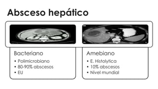
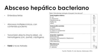
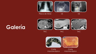
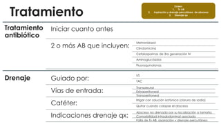
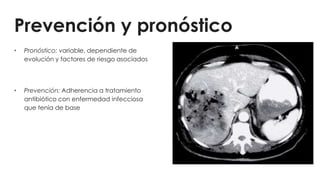
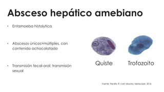
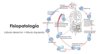
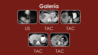
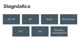
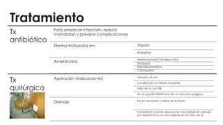
Absceso hepático
amebiano y bacteriano
Leal Lam Sara Li
482
Gastroenterología
UNIVERSIDAD AUTÓNOMA DE BAJA CALIFORNIA
Escuela de Ciencias de la Salud
Unidad Valle de las Palmas
Absceso hepático
Bacteriano
• Polimicrobiano
• 80-90% abscesos
• EU
Amebiano
• E. Histolytica
• 10% abscesos
• Nivel mundial
Absceso hepático bacteriano
• Enterobacterias
• Abscesos múltiples>únicos, con
contenido purulento
• Transmisión directa (tracto biliar), vía
hematógena (circ. portal), criptógena
• Fatal si no es tratada
Fuente: Peralta, R. Liver abscess. Medscape. 2016.
Epidemiología
• El tipo más común de los abscesos
viscerales
• Hombres ≥ mujeres, ≥60 años
• FR: DM, enfermedad hepatobiliar o
pancreática de base, trasplante de
hígado, factores geográficos
• Mortalidad 5-30%
Fuente: Davis J. Pyogenic liver abscess. Uptodate. 2017.
Fisiopatología
Lóbulo derecho>lóbulo izquierdo
Manifestaciones clínicas
Síntomas
•Fiebre
•Dolor abdominal
•Náusea, vómito
•Anorexia
•Malestar
Signos
•Dolor en hipocondrio derecho
•Hepatomegalia
•Ictericia
•Hemidiafragma elevado
Complicaciones
Pleuropulmonar
Pleuritis
Derrame pleural
Empiema
Fístula
broncohepática
Intraabdominal
Absceso
subfrénico
Rotura de
absceso hacia
cavidad
peritoneal
Otros
Encefalopatía
Sepsis
Síndrome de falla
multiorgánica
Diagnóstico
HC+EF BH Ultrasonido
TAC Cultivo
Galería
Placa de tórax US US
TAC TAC TAC
Corte
macroscópico
Corte
macroscópico
Tratamiento
Tratamiento
antibiótico
Iniciar cuanto antes
2 o más AB que incluyen:
Metronidazol
Clindamicina
Cefalosporinas de 3ra generación IV
Aminoglucósidos
Fluoroquinolonas
Drenaje Guiado por:
US
TAC
Vías de entrada:
Transpleural
Extraperitoneal
Transperitoneal
Catéter:
Irrigar con solución isotónica (cloruro de sodio)
Quitar cuando colapse el absceso
Indicaciones drenaje qx:
Absceso no drenado por su localización o tamaño
Comorbilidad intraabdominal asociada
Falla de Tx AB, aspiración y drenaje percutáneo
Orden:
1. Tx AB
2. Aspiración y drenaje percutáneo de absceso
3. Drenaje qx
Prevención y pronóstico
• Pronóstico: variable, dependiente de
evolución y factores de riesgo asociados
• Prevención: Adherencia a tratamiento
antibiótico con enfermedad infecciosa
que tenía de base
Absceso hepático amebiano
• Entamoeba histolytica
• Abscesos únicos>múltiples, con
contenido achocolatado
• Transmisión fecal-oral; transmisión
sexual
Quiste Trofozoito
Fuente: Peralta, R. Liver abscess. Medscape. 2016.
Epidemiología
• Afecta más a hombres, 30-50 años,
inmigrantes o viajeros
• RARO (10% abscesos hepáticos)
• FR: áreas endémicas (países en
desarrollo); hacinamiento, mala higiene,
muchedumbre, inmunosupresión
Fisiopatología
Lóbulo derecho > lóbulo izquierdo
Manifestaciones clínicas
Dolor
abdominal
Fiebre Hepatomegalia
Náusea/vómito Diarrea
Síntomas
pulmonares
Inicio de síntomas :
2-5 meses después
de infección
Galería
US TAC TAC
TAC TAC
Complicaciones
Pleuropulmonares
Efusión serosa
Ruptura hacia
cavidad torácica
(empiema)
Vía hematógena
Fístula
broncopleural
Esputo con
aspecto de “pasta
de anchoas”
Trofozoitos en
esputo
Absceso puede
curar
espontáneamente
Afectación
cardíaca
Asociado a gran
mortalidad
Ruptura de
absceso en lóbulo
izquierdo
Ruptura
intraperitoneal
2-7% pacientes
Lóbulo
izquierdo>lóbulo
derecho
Otros
Sobreinfección
bacteriana
Pseudoaneurisma
de arteria
hepática
Ruptura hacia
estómago o
mediastino
Diagnóstico
HC+EF BH ELISA Ultrasonido
TAC RM
Estudios
coprológicos
Tratamiento
Tx
antibiótico
Para erradicar infección, reducir
mortalidad y prevenir complicaciones
Elimina trofozoitos en: Hígado
Intestino
Amebicidas:
Metronidazol (1ra elección)
Tinidazol
Dehidroemetina
Cloroquina
Tx
quirúrgico
Aspiración (indicaciones) Tamaño >5 cm
Localización en lóbulo izquierdo
Falla de Tx con AB
No se puede diferenciar de un absceso piógeno
Drenaje No es necesario y debe ser evitado
Considerar cuando absceso es inaccesible en drenaje
por aspiración o no hay mejoría en 5-7 días de Tx
Prevención y pronóstico
Prevención:
• No hay fármacos profilácticos
• Control adecuado de excretas
• Hervir agua
• Lavado de manos
• Desinfección
Pronóstico: Generalmente bueno
-Resolución radiológica: 12 meses (promedio)
Bibliografía
Texto:
1. Peralta, R. Liver abscess. Medscape. 2016
2. Nickloes T. Pyogenic hepatic abscesses. Medscape. 2016.
3. Arora K. Liver and intrahepatic bile ducts-nont umor: Infectious (non-viral) disorders: Bacterial infections. Pathology Outlines. 2012.
Imágenes.
1. Peralta, R. Liver abscess. Medscape. 2016
2. Nickloes T. Pyogenic hepatic abscesses. Medscape. 2016.
3. Fang D., Shu D. Entamoeba histolytica liver abscess. CMAJ. 2010 Nov 9; 182(16): 1758. doi: 10.1503/cmaj.091926.

Absceso hepático amebiano y bacteriano

  • 1.
    Absceso hepático amebiano ybacteriano Leal Lam Sara Li 482 Gastroenterología UNIVERSIDAD AUTÓNOMA DE BAJA CALIFORNIA Escuela de Ciencias de la Salud Unidad Valle de las Palmas
  • 2.
    Absceso hepático Bacteriano • Polimicrobiano •80-90% abscesos • EU Amebiano • E. Histolytica • 10% abscesos • Nivel mundial
  • 3.
    Absceso hepático bacteriano •Enterobacterias • Abscesos múltiples>únicos, con contenido purulento • Transmisión directa (tracto biliar), vía hematógena (circ. portal), criptógena • Fatal si no es tratada Fuente: Peralta, R. Liver abscess. Medscape. 2016.
  • 4.
    Epidemiología • El tipomás común de los abscesos viscerales • Hombres ≥ mujeres, ≥60 años • FR: DM, enfermedad hepatobiliar o pancreática de base, trasplante de hígado, factores geográficos • Mortalidad 5-30% Fuente: Davis J. Pyogenic liver abscess. Uptodate. 2017.
  • 5.
  • 6.
    Manifestaciones clínicas Síntomas •Fiebre •Dolor abdominal •Náusea,vómito •Anorexia •Malestar Signos •Dolor en hipocondrio derecho •Hepatomegalia •Ictericia •Hemidiafragma elevado
  • 7.
  • 8.
  • 9.
    Galería Placa de tóraxUS US TAC TAC TAC Corte macroscópico Corte macroscópico
  • 10.
    Tratamiento Tratamiento antibiótico Iniciar cuanto antes 2o más AB que incluyen: Metronidazol Clindamicina Cefalosporinas de 3ra generación IV Aminoglucósidos Fluoroquinolonas Drenaje Guiado por: US TAC Vías de entrada: Transpleural Extraperitoneal Transperitoneal Catéter: Irrigar con solución isotónica (cloruro de sodio) Quitar cuando colapse el absceso Indicaciones drenaje qx: Absceso no drenado por su localización o tamaño Comorbilidad intraabdominal asociada Falla de Tx AB, aspiración y drenaje percutáneo Orden: 1. Tx AB 2. Aspiración y drenaje percutáneo de absceso 3. Drenaje qx
  • 11.
    Prevención y pronóstico •Pronóstico: variable, dependiente de evolución y factores de riesgo asociados • Prevención: Adherencia a tratamiento antibiótico con enfermedad infecciosa que tenía de base
  • 12.
    Absceso hepático amebiano •Entamoeba histolytica • Abscesos únicos>múltiples, con contenido achocolatado • Transmisión fecal-oral; transmisión sexual Quiste Trofozoito Fuente: Peralta, R. Liver abscess. Medscape. 2016.
  • 13.
    Epidemiología • Afecta mása hombres, 30-50 años, inmigrantes o viajeros • RARO (10% abscesos hepáticos) • FR: áreas endémicas (países en desarrollo); hacinamiento, mala higiene, muchedumbre, inmunosupresión
  • 14.
  • 15.
    Manifestaciones clínicas Dolor abdominal Fiebre Hepatomegalia Náusea/vómitoDiarrea Síntomas pulmonares Inicio de síntomas : 2-5 meses después de infección
  • 16.
  • 17.
    Complicaciones Pleuropulmonares Efusión serosa Ruptura hacia cavidadtorácica (empiema) Vía hematógena Fístula broncopleural Esputo con aspecto de “pasta de anchoas” Trofozoitos en esputo Absceso puede curar espontáneamente Afectación cardíaca Asociado a gran mortalidad Ruptura de absceso en lóbulo izquierdo Ruptura intraperitoneal 2-7% pacientes Lóbulo izquierdo>lóbulo derecho Otros Sobreinfección bacteriana Pseudoaneurisma de arteria hepática Ruptura hacia estómago o mediastino
  • 18.
    Diagnóstico HC+EF BH ELISAUltrasonido TAC RM Estudios coprológicos
  • 19.
    Tratamiento Tx antibiótico Para erradicar infección,reducir mortalidad y prevenir complicaciones Elimina trofozoitos en: Hígado Intestino Amebicidas: Metronidazol (1ra elección) Tinidazol Dehidroemetina Cloroquina Tx quirúrgico Aspiración (indicaciones) Tamaño >5 cm Localización en lóbulo izquierdo Falla de Tx con AB No se puede diferenciar de un absceso piógeno Drenaje No es necesario y debe ser evitado Considerar cuando absceso es inaccesible en drenaje por aspiración o no hay mejoría en 5-7 días de Tx
  • 20.
    Prevención y pronóstico Prevención: •No hay fármacos profilácticos • Control adecuado de excretas • Hervir agua • Lavado de manos • Desinfección Pronóstico: Generalmente bueno -Resolución radiológica: 12 meses (promedio)
  • 21.
    Bibliografía Texto: 1. Peralta, R.Liver abscess. Medscape. 2016 2. Nickloes T. Pyogenic hepatic abscesses. Medscape. 2016. 3. Arora K. Liver and intrahepatic bile ducts-nont umor: Infectious (non-viral) disorders: Bacterial infections. Pathology Outlines. 2012. Imágenes. 1. Peralta, R. Liver abscess. Medscape. 2016 2. Nickloes T. Pyogenic hepatic abscesses. Medscape. 2016. 3. Fang D., Shu D. Entamoeba histolytica liver abscess. CMAJ. 2010 Nov 9; 182(16): 1758. doi: 10.1503/cmaj.091926.

Notas del editor

  • #5 Untreated, pyogenic liver abscess remains uniformly fatal. With timely administration of antibiotics and drainage procedures, mortality currently occurs in 5-30% of cases. The most common causes of death include sepsis, multiorgan failure, and hepatic failure.
  • #6 Pyogenic or bacterial abscess may be caused by several factors and is classified by the route of entry of the organisms. Infections may arise from the biliary tract, portal vein and hepatic artery or by direct extension. Infections arising from the biliary tract are the most common and result in 30% to 50% of the total number of pyogenic abscesses. The resultant cholangitis leads to liver abscesses, which are frequently multiple. Biliary obstruction is commonly present from causes such as choledocholithiasis and benign or malignant strictures. Other causes of cholangitis include iatrogenic intervention from endoscopic retrograde cholangiopancreatography (ERCP) or percutaneous transhepatic procedures. Another common route of entry of infection is the portal vein. Conditions such as complicated diverticular disease, appendicitis, peritonitis and pancreatitis may cause portal vein pyaemia, resulting in pyogenic liver abscesses. Septicemia from any cause may also give rise to multiple liver abscesses via dissemination from the hepatic artery. These account for 5 to 15% of pyogenic liver abscesses. Common causes include bacterial endocarditis, pneumonia and intravenous drug abuse. Other causes of liver abscesses include complicated liver trauma from blunt or penetrating causes, or by direct extension from other conditions such as empyema of the gall bladder. In a number of cases, the cause is not obvious. The infecting organism varies according to the site of entry. In biliary or portal vein sepsis, the organisms are enteric and usually polymicrobial. Staphylococcus aureus is evident in 20% of cases and is confirmed predominantly from haematogenous spread.
  • #7 Presentación gradual (semanas).
  • #9 Antecedente de infección en tracto GI (peritonitis, diverticulitis, apendicitis, colangitis, etc). The clinical presentation of liver abscess is insidious; many patients have symptoms for weeks before presentation. Fever and right-upper-quadrant (RUQ) pain are the most common complaints. Pain is reported in as many as 80% of patients and may be associated with pleuritic chest pain or right shoulder pain. Symptoms are often misdiagnosed as acute cholecystitis. Fever occurs in 87-100% of patients and is usually associated with chills and malaise. [9, 21]  Anorexia, weight loss, and mental confusion are also common symptoms. A complete blood count (CBC) should be obtained. Anemia is observed in 50-80% of patients. [8, 9] Leukocytosis of more than 10,000/μL is observed in 75-96% of patients. [8, 9] Bands of more than 10% are observed in 40% of patients. The erythrocyte sedimentation rate (ESR) is commonly elevated. Liver function tests are helpful. An elevated alkaline phosphatase level [4] is observed in 95-100% of patients. [8, 9] An elevated serum aspartate aminotransferase level, an elevated serum alanine aminotransferase level, or both are observed in 48-60% of patients. An elevated bilirubin level [14] is observed in 28-73% of patients. [8, 9] A decreased albumin level (<3 g/dL) and an increased globulin value (>3 g/dL) are frequently observed. The prothrombin time (PT) is elevated in 71-87% of patients.  Real-time ultrasonography findings are 80-100% sensitive. A round or oval hypoechoic mass is consistent with pyogenic abscess. CT has become the imaging study of choice for detecting liver lesions. Pyogenic liver abscesses are not enhanced on images after intravenous contrast administration. Triphasic CT scanning with arterial and portal venous phases helps to define the proximity of the abscess to the major branches of the portal and hepatic veins. Findings have sensitivity similar to that of ultrasonography, but they lack specificity. Diagnostic aspiration is performed under ultrasonographic or CT guidance [9, 16] and is usually followed by drainage catheter placement. The aspirate is sent for culture and cytology.
  • #13 Entamoeba histolytica (90%); Entamoeba dispar (10%) Worldwide, approximately 40-50 million people are infected annually, with the majority of infections occurring in developing countries. The prevalence of infection is higher than 5-10% in endemic areas [4] and sometimes as high as 55%. [5] The highest prevalence is found in developing countries in the tropics, particularly in Mexico, India, Central and South America, and tropical areas of Asia and Africa. Infection with E histolytica ranks second worldwide among parasitic causes of death, following malaria. Annually, 40,000-100,000 deaths are caused by infection with E histolytica. Per year, a 10% risk of developing symptomatic invasive amebiasis exists after the acquisition of a pathogenic strain.
  • #15 E histolytica exists in 2 forms. The cyst stage is the infective form, and the trophozoite stage causes invasive disease. People who chronically carry E histolytica shed cysts in their feces; these cysts are transmitted primarily by food and water contamination. Rare cases of transmission via oral and anal sex or direct colonic inoculation through colonic irrigation devices have occurred. Cysts are resistant to gastric acid, but the wall is broken down by trypsin in the small intestine. Trophozoites are released and colonize the cecum. To initiate symptomatic infection, E histolytica trophozoites present in the lumen must adhere to the underlying mucosa and penetrate the mucosal layer. Liver involvement occurs following invasion of E histolytica into mesenteric venules. Amebae then enter the portal circulation and travel to the liver where they typically form large abscesses. The abscess contains acellular proteinaceous debris, which is thought to be a consequence of induced apoptosis [2] and is surrounded by a rim of amebic trophozoites invading the tissue. The right lobe of the liver is more commonly affected than the left lobe. This has been attributed to the fact that the right lobe portal laminar blood flow is supplied predominantly by the superior mesenteric vein, whereas the left lobe portal blood flow is supplied by the splenic vein.
  • #16 Presentación aguda (<14 días). Síntomas inician 8-12 semanas después de un viaje; 95% manifiesta síntomas en un periodo de 5 meses posterior a un viaje hacia una zona endémica. -Dolor abdominal: 90-93% de los pacientes. Dolor tipo sordo, constante, en cuadrante superior derecho que puede irradiar a hombro o escápula derecha. Aumenta con respiración profunda, tos, al caminar o al recargarse sobre su lado derecho. -Fiebre: 87-100% de los pacientes. -Hepatomegalia: En algunos casos (variable). A la EF hay aumento de tamaño con dolor al aplicar presión sobre área hepática, debajo de las costillas o en los espacios intercostales; predomina en lóbulo derecho>lóbulo izquierdo (si se presenta en este último, puede haber sensibilidad en epigastrio). -N/V: hasta en el 85% de los Px. Pérdida de peso en hasta el 64% de los Px. -Diarrea: Menos de un tercio de los Px. Algunos describen cuadros de disentería. -Síntomas pulmonares: Menos del 30% de los Px. Los más frecuentes son tos y dolor torácico, que se dan cuando hay complicación a cavidad torácica. Si hay producción de esputo color café (“pasta de anchoas”) sin olor, indica fístula broncopleural. A la EF, hay matidez a la percusión o crepitantes en lóbulo inferior del pulmón derecho y tos no productiva, con disminución de los ruidos respiratorios y puede haber roce pleural. Hay ictericia en <10% de los Px, y cuando hay múltiples abscesos o uno muy grande que comprima el tracto biliar.
  • #18 Signs of complications include the following: Signs of peritoneal irritation, such as rebound tenderness, guarding, and absence of bowel sounds, are present when the abscess ruptures into the peritoneal cavity. Peritonitis occurs in 2-7% of cases. Pericardial friction rub can be audible when the abscess extends into the pericardium. This sign is associated with a very high mortality. Signs of pleural effusion are present when the abscess ruptures into the pleural cavity.