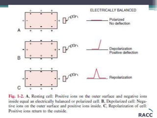
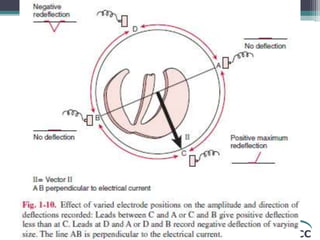
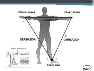
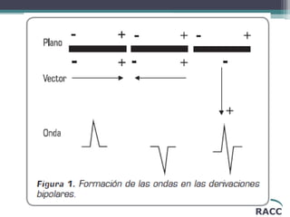
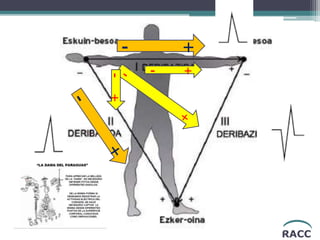
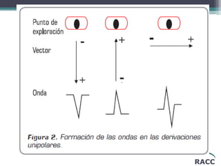
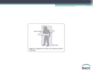
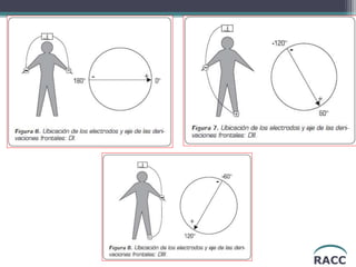
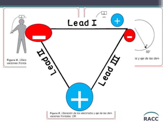
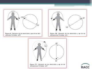
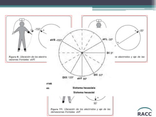
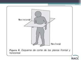
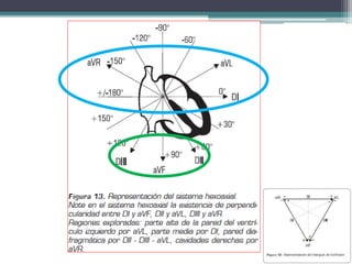
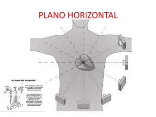
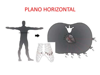
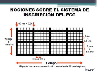
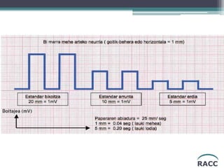
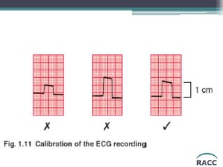
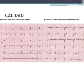
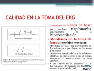
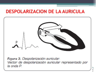
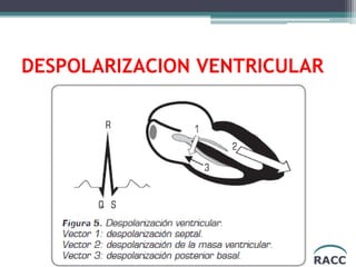
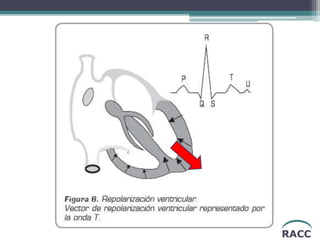
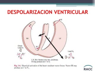
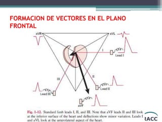
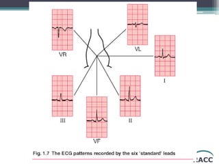
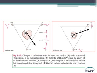
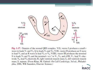
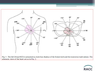
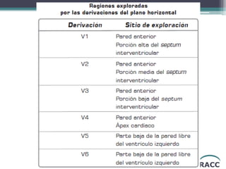
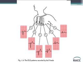
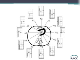
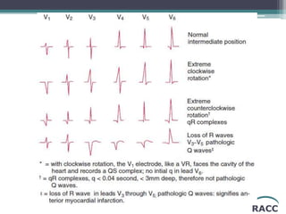
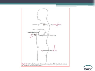
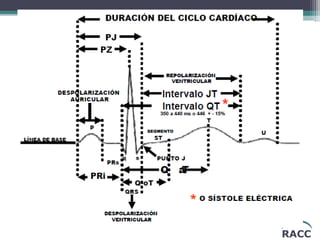
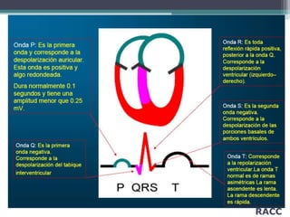
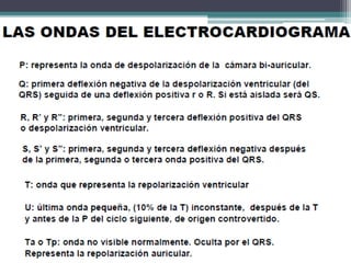
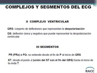
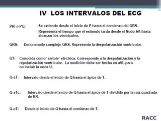
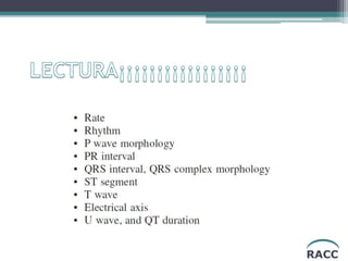
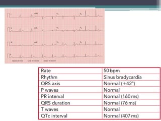
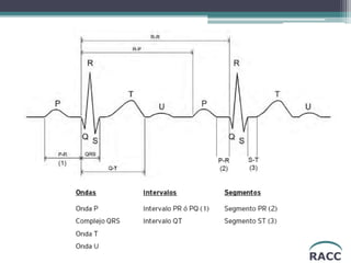
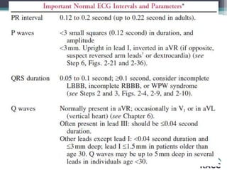
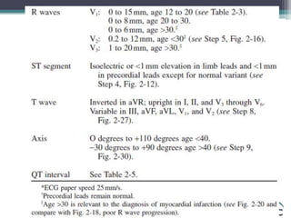
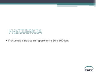
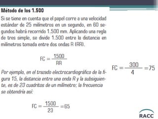
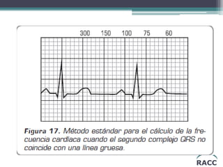
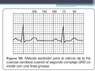
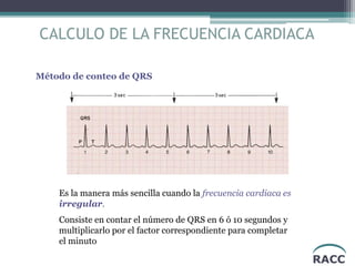
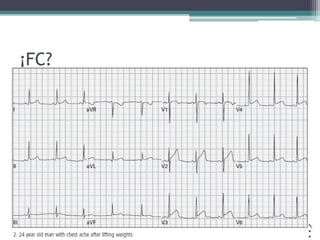
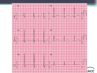
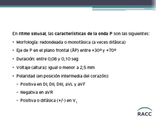
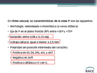
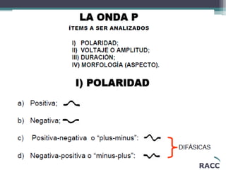
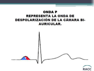
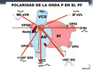
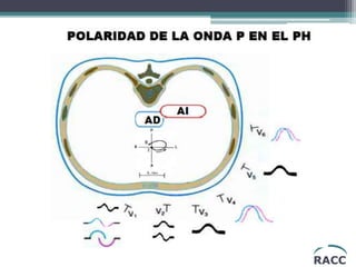
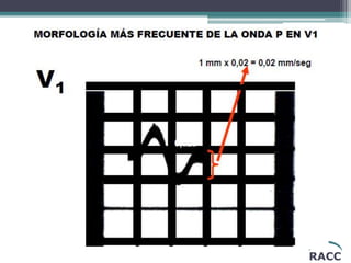
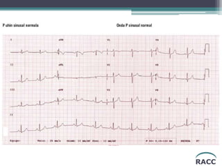
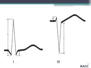
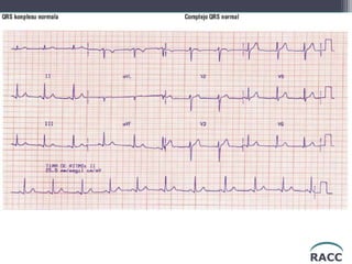
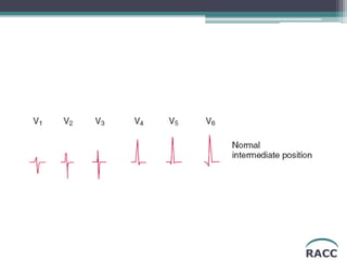
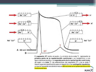
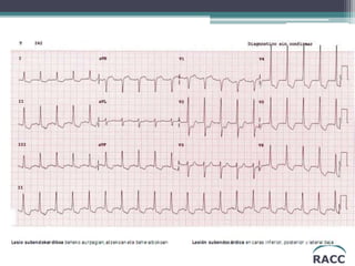
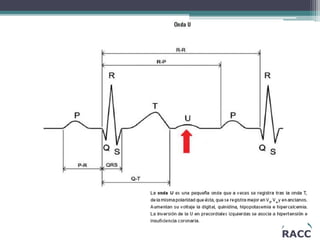
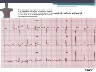
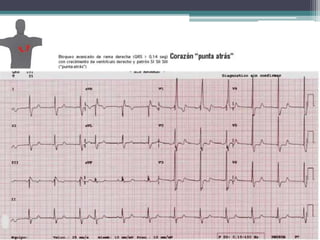
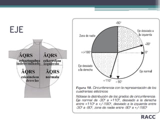
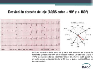
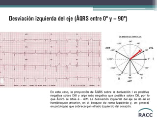
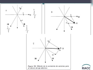
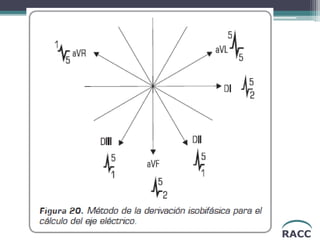
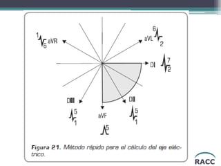
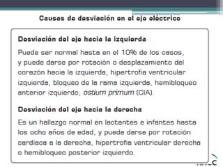
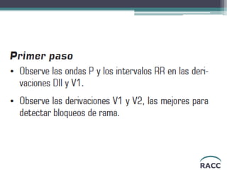
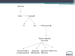
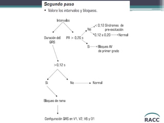
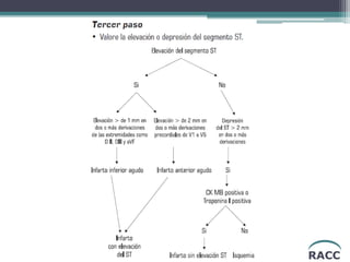
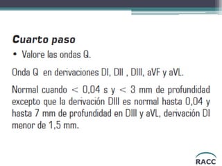
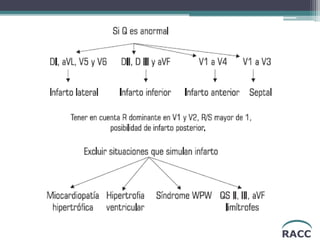
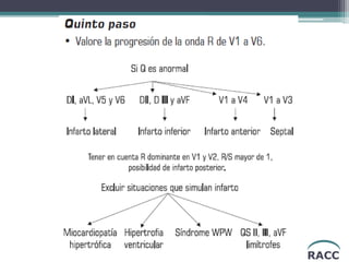
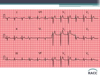
29. RACC
CALIDAD EN LA TOMA DEL EKG
• Alteraciones en la línea de base”
por cambios respiratorios,
especialmente en la
hiperventilación.
• Mordiscos en la línea de
base por temblor muscular.
• “Pérdidas de foco” por movimientos de
los miembros o por llanto, en los casos
pediátricos
• “Líneas en empalizada” por interferencia
eléctrica, la cual se debe a que el equipo
comparte el tomacorriente con otro
aparato.
• • Los “saltos en la secuencia” o “el
borramiento” del trazado por la pérdida
de contacto de un electrodo
59. RACC
CALCULO DE LA FRECUENCIA CARDIACA
Método de conteo de QRS
Es la manera más sencilla cuando la frecuencia cardiaca es
irregular.
Consiste en contar el número de QRS en 6 ó 10 segundos y
multiplicarlo por el factor correspondiente para completar
el minuto
68. RACC
MENOR DE 2 ½
CUADRADITOS… PARA
ARRIBA Y PARA ABAJO
Positiva o Bifásica en V1...
Positivaen resto
de plano horizontal
POSITIVAEN
TODAS LAS
FRONTALES, excepto en
aVR (negativa)
72. RACC
MENOR DE 2 ½
CUADRADITOS… PARA
ARRIBA Y PARA ABAJO
Positiva o Bifásica en V1...
Positivaen resto
de plano horizontal
POSITIVAEN
TODAS LAS
FRONTALES, excepto en
aVR (negativa)La Derecha es de Altura,
pero la Izquierda Durara
siempre
Lead I connects the two arms and is formed by connecting L to the positive terminal and R to the negative terminal of the galvanometer; thus, I = aVL = aVR. Lead I looks at the heart from the left, inferior to lead aVL, the lead of the left shoulder (arm), and displays the electrical tracing produced by a combination of the right arm and left arm electrodes.
The right leg electrode is an earth (or ground) and minimizes interference.
• Lead II looks at the heart from a position to the left of the left groin, foot lead F (see Fig. 1-12).
• Lead III looks at the heart from a position to the right of the left groin, foot lead F. Thus, leads II, III, and aVF look at the inferior surface of the heart from different angles, and they usually show some similarities.
Lead III is the most unreliable of the leads II, III, and aVF. Thus, many errors are made from the observation of the QRS and T wave in lead
VECTOR III• Activation of the posterobasal right and left ventricular free walls and
the basal right septal mass, including the crista supraventricularis, represents
vector III.
• The resultant force is directed to the right, is small in magnitude, and
may record a small S wave in V5 and V6 and a terminal r′ wave in
lead V1 or V2; thus, an Rsr′ pattern in V1 may occur in normal
individuals.
ECG que exibe un complejo QRS de anchura (0,09 seg) y voltaje normales (voltaje de DI+DII+DIII = 32 mm, voltaje de V1 + V6= 25 mm), con eje eléctrico (ÂQRS) a +80º, características propias de un complejo QRS normal.