
Recomendados
Más contenido relacionado
La actualidad más candente
La actualidad más candente (20)
Similar a Fracturas de Lefort
Similar a Fracturas de Lefort (20)
Último
Último (20)
Fracturas de Lefort
- 1. FRACTURAS DE LEFORT BENEMÉRITA UNIVERSIDAD AUTÓNOMA DE PUEBLA FACULTAD DE MEDICINA EMMANUEL NUÑEZ SALDAÑA
- 2. CLINICA • ASIMETRÍAS EN EL MEDIO FACIAL • DOLOR A LA PRESIÓN • HEMATOMA PERIORBITARIO BILATERAL O EN ANTIFAZ • EPISTAXIS • EQUIMOSIS CONJUNTIVAL • AUMENTO DE LA LONGITUD DEL TERCIO MEDIO FACIAL (CARA LARGA, CARA DE PLATO). • MALA OCLUSIÓN.
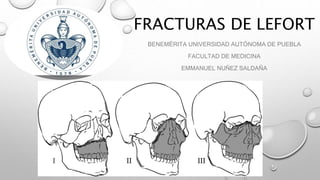
- 4. Fractura le fort I o de guerin o transversal de maxilar superior • Esta fractura compromete el maxilar superior, provocando una disyunción de éste. El rasgo de fractura recorre en una dirección ántero posterior: la espina nasal anterior y tabique nasal, cara externa del maxilar superior sobre los ápices dentarios, pared anterior y posterior del seno maxilar, proceso cigomático-alveolar y procesos pterigoides.
- 5. • CLÍNICAMENTE SE OBSERVA DOLOR Y EDEMA A NIVEL DEL LABIO SUPERIOR. EQUIMOSIS EN EL FONDO DEL VESTÍBULO BUCAL SUPERIOR Y EN LA REGIÓN PALATINA POSTERIOR • ENFISEMA EN LOS TEJIDOS BLANDOS DE LAS REGIONES GENIANA Y CIGOMÁTICA, POR EL COMPROMISO DE LAS PAREDES DEL SENO MAXILAR. • UN SIGNO CARACTERÍSTICO DE ESTA FRACTURA ES EL MOVIMIENTO EN BLOQUE DEL MAXILAR SUPERIOR, QUE SE MANIFIESTA MEDIANTE UNA MANIOBRA QUE CONSISTE EN MANIPULAR EL MAXILAR TOMÁNDOLO DESDE LAS PIEZAS DENTARIAS, PARA OBSERVAR LA MOVILIDAD DE ÉSTE.
- 6. •Fractura le fort II o piramidal: • La línea de fractura discurre por la raíz nasal, hueso lacrimal, reborde infraorbitario y por la pared del maxilar hasta la apófisis pteriogides.
- 7. • Fractura le fort III o disyunción cráneo facial • Raíz nasal, hueso lacrimal, apófisis frontal del hueso malar, pared lateral y posterior del maxilar hasta apófisis pterigoides.