Angiofibroma nasofaringeo juvenil
•Descargar como PPTX, PDF•
5 recomendaciones•4,005 vistas

Angiofibroma, nasoangiofibroma, nasofaringeo, juvenil

Recomendados
Más contenido relacionado
La actualidad más candente
La actualidad más candente (20)
Similar a Angiofibroma nasofaringeo juvenil
Similar a Angiofibroma nasofaringeo juvenil (20)
Más de Angel Castro Urquizo
Más de Angel Castro Urquizo (20)
Último
Último (20)
Angiofibroma nasofaringeo juvenil
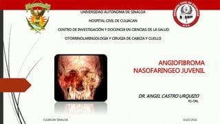
- 1. ANGIOFIBROMA NASOFARINGEO JUVENIL UNIVERSIDAD AUTONOMA DE SINALOA HOSPITAL CIVIL DE CULIACAN CENTRO DE INVESTIGACIÓN Y DOCENCIA EN CIENCIAS DE LA SALUD OTORRINOLARINGOLOGIA Y CIRUGIA DE CABEZA Y CUELLO DR. ANGEL CASTRO URQUIZO R1 ORL CULIACAN SINALOA JULIO 2016
- 2. Introducción Masa vascular mas común en nariz Neoplasia benigna nasofaríngea mas común Exclusivo de hombres adolescentes Hipócrates 500 BC Friedberg 1940 la nombro • Nasofibroma juvenil • Fibroma nasofaríngeo • Nasoangiofibroma juvenil • etc
- 3. Epidemiologia 0,5% de todos los tumores de cabeza y cuello Exclusivo en hombres Mujeres someterse a pruebas genéticas Segunda década 7-19 años >25 años muy raro
- 4. Etiopatogenia Inserción posterior cornete medio Borde superior del agujero esfenopalatino Receptores hormonales y VEFG Células paraganglionicas de ramas terminales arteria maxilar Remanentes vasculares
- 5. Etiopatogenia Comienza en foramen esfenopalatino Bilobulados Forma de mancuerna Una porción en nasofaringe La otra hacia la fosa pterigopalatina Llena cavidad nasal unilateral
- 6. Clínica Obstrucción nasal (90%) Epistaxis (70%) Cefalea Edema facial Rinorrea unilateral Anosmia Rinolalia Otalgia Plenitud
- 7. Clínica Masa nasal (80%) Masa orbitaria (20%) Proptosis Otitis serosa Edema cigomático Trismus Alteración visuales
- 9. Diagnostico diferencial Other causes of nasal obstruction, (eg, nasal polyps, antrochoanal polyp,teratoma, encephalocele, dermoids, inverting papilloma, rhabdomyosarcoma, squamous cell carcinoma) Other causes of epistaxis, systemic or local Other causes of proptosis or orbital swellings
- 10. Diagnostico
- 18. Diagnostico Histopatología Sésil Lobulado Rojo, rosáceo Puede ser polipoide o pedunculado Encapsulado, vascular, estroma fibroso con fibras de colágeno Abundantes mastocito Inmunohistoquimica Vimentina
- 20. Estadios de Fisch I.- Cavidad nasal II.- Fosa pterigomaxilar, senos paranasales con destrucción ósea III.- Fosa infratemporal Orbita Area paraselar IV.- Seno cavernoso Quiasma óptico Fosa pituitaria
- 21. Tratamiento Flutamida estadio I y II Reduce 45% Radioterapia coadyuvante preoperatorio, extensión intracraneal 30 Gy en 15 sesiones por 3 semanas Quimioterapia: solo en recurrencias Cuando no se pueda operar o administrar radioterapia
- 22. Tratamiento quirúrgico Fisch I , II rinotomia lateral, transpalatinono, transmaxilar, esfenoetmoidal I Abordaje endoscópico transnasal II Enfoque combinado: trans nasal – infratemporal III Abordaje extra e intracraneal
- 26. Preoperatorio Embolizacion transarterial Gelfoam, polivinil, dextran, ivalon, terbal, etc
- 28. Objetivo Evaluar factibilidad de la resección de angiofibroma (JNA) con ablación por radiofrecuencia endoscópica (RFA) Comparar resultados con resección tradicional endoscópica (TE) Diseño: Serie de casos
- 29. Ablación por radiofrecuencia bipolar Campo eléctrico Desnaturaliza proteínas Ablación de tejidos Propiedad coagulativa
- 30. Métodos Pacientes con JNA sometidos a cirugía endoscópica 2000 – 2014 Factores demográficos, estancia hospitalaria, detalles, resultados operatorios y posoperatorios. 29 pacientes Excluidos resto de abordajes y los de etapa I Tiempo operatorio, perdidas sanguíneas, transfusión Estancia hospitalaria, recurrencias
- 31. Resultados 16 sometidos a TE 13 RFA Edades promedio TE 15 @ RFA 15,5 @
- 34. Discusión Falta de ensayos clínicos controlados por lo raro de la enfermedad RFA es técnica alternativa y factible Sin diferencias en tiempo quirúrgico, sangrado, tiempo hospitalario
- 35. Limitaciones Grupos pequeños Falta de aleatorizar la modalidad Cirugías con RFA fueron realizadas mas recientemente Periodos de seguimiento cortos
- 36. Conclusión No existieron ventajas estadísticamente significativas con RFA comparado con TE. Sin diferencias en cuanto a tiempo quirúrgico, estancia hospitalaria, perdidas sanguíneas, tasas de recurrencias. Alternativa quirúrgica
Notas del editor
- 2530 2542
- Hipocrates la describió En adultos:.: regresión espontanea después de los 25 años
- BENIGNO PERO CON CARACTERISTICAS MALIGNAS CRECE LENTAMENTE, INVASIVIDAD LOCAL PUEDE INVADIR INTRACRANEAL NO METS 5% en mujeres
- PARED POSTERO LATERAL CAVIDA NASALL. Cerca del foramen esfenopalatino Altos niveles ed receptores ghormonales, factor de recimmiento vacular endotelial REAMNENTES : REGRESION INCOMPLETA,
- Desviacion septal hacia el otro lado, invasiona fosa nasal y seno maxilar Crecimiento superior hacia el seno esfenoidal Inclusive puede erosionar Seno cavernoso puede ser invadido Proptotiss ocular---- atrofia óptica
- Obstruccion… síntoma mas comun e inicial Epistaxis. Unukateral, recurrente, Deformidad de la mejilla, Edema de paladar
- Trismus por invsion a la fosa infratemporal Afectacion N. II
- Juvenile angiofibroma. On endoscopy, the lesion typically appears as a polypoid hypervascularized mass bulging from the lateral wall behind the middle turbinate, which is laterally compressed. The choana is completely obstructed.
- Biopsia– rara vez justificada por riesgos de sangrado
- Coronal CT scan of the lesion filling the left nasal cavity and ethmoid sinuses, blocking the maxillary sinus and deviating the nasal septum to the right side.
- Axial CT scan of lesion involving the right nasal cavity and paranasal sinuses. Courtesy of J Otolaryngol 1999;28:145.
- Coronal MRI scan showing extension of the lesion to the cavernous sinus. Courtesy of J Otolaryngol 1999;28:145.
- Juvenile angiofibroma on coronal contrast-enhanced magnetic resonance (MR) images obtained before and after endoscopic resection. A, Pretreatment image shows encroachment of both the floor and lateral wall of the left sphenoid sinus and infratemporal fossa. Intracranial extension is demonstrated in close proximity to the superior orbital fissure (arrows). B, MR image obtained after surgical resection shows solid tissue (arrows) along the lateral sphenoid sinus wall; the lack of contrast enhancement suggests residual scar tissue.
- Juvenile angiofibroma on axial T2-weighted, spin-echo magnetic resonance image. The lesion invades the left orbital apex (OA), the lateral wall of the left sphenoid sinus (white arrows) is completelydestroyed, and the internal carotid artery (ICA) is encased. On the right side, a bony barrier (arrowheads) still separates the lesion from the internal carotid artery.
- Carotida Externa maxilar interna tambien pueden contribuir faringea ascendete, vidiana Rara vez ramas de la carotida interna Angiogram depicting angiofibroma before y after embolization. Courtesy of J Otolaryngol 1999;28:145.
- 3.- sin compromiso intracraneal Selar.. Silla turca
- Bloqueador de receptores testosterona Han reportado … RADIOTERAPIA: 80% de contorl
- Abordaje transpalatino
- 48 horas antes de la cirugía Recurrencia alrededor de 45% de los casos
- Departamen orl pensiolvania
- RFA: crea calor en punta de electroldo. Provoca necrosis coagulativa. Desnatuarliza proterina, rompe mebranas celulares.
- 29 cumplieron los criterios de inclusion
- Sin tanta diferencia entre el tipo de procedimiento y el estadio There was no significant difference in mean 6 SD age between the 2 cohorts (TE, 14.8 6 2.8 years; RFA, 15.7 6 3.3 years; P = .41). Following elimination of the 6 stage 1 patients (as previously described), the distribution of UPMC classification scores between the groups was not statistically different (P = .79). There was also no significant difference in the distribution of type of procedure performed at the given hospitals (P = .99;
- Sin diferencias significativas entre ambos grupos respecto al tiempo operativo There was no significant difference in operative time between the 2 groups (TE, 6.3 6 3.4 hours; RFA, 7.8 6 4.3 hours; P = .29; Table 2). The average blood loss and need for blood transfusion were also not statistically different between the 2 groups (TE, 375 mL; RFA, 500 mL; P = .27; TE, 31.3%; RFA, 46.2%; P = .47, respectively). Length of hospitalization was not significantly different between groups (TE, 3.8 6 2.3 days; RFA, 4.3 6 1.1 days; P = .46). Finally, there was no significant difference in recurrence rates (TE, 25%; RFA, 23%; P = .99). The average follow-up period was 1.7 years (range, 1 month to 5 years) in the RFA group versus 2.5 years (range, 1 month to 7 years) in the TE group (P = .23). There were no significant differences between RFA and TE for intraoperative and postoperative outcomes after adjusting for age, UPMC classification, and center.
- seosgos