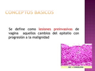
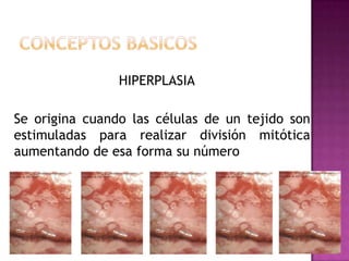
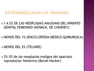
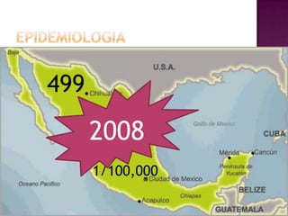
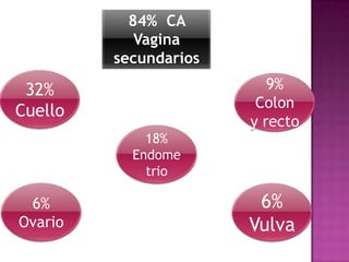
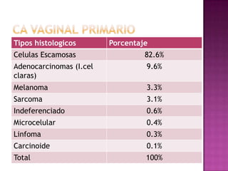
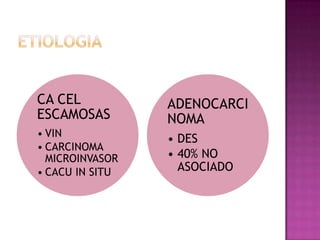
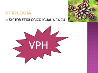
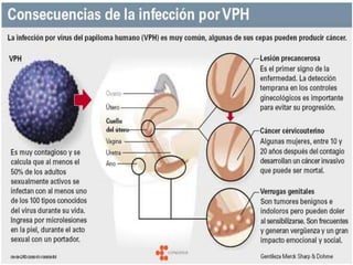
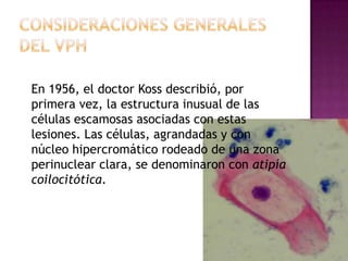
Este documento describe lesiones preinvasivas y canceres de vagina. Define hiperplasia, metaplasia y displasia como lesiones preinvasivas que pueden progresar a cáncer. Explica que la mayoría de cánceres de vagina son metastáticos de cáncer de cuello uterino o vulva, y que los virus del papiloma humano (VPH) causan alrededor del 90% de cánceres de vagina. Resume los síntomas, tipos histológicos, estadios y tratamientos de cáncer de vagina
Vaginal Cancer, cáncer de vagina
Surgery and radiation therapy outcomes
Etapificación, epidemiología, resultados con braquiterapia, radioterapia externa.
Se define como lesiones premalignas a todas aquellas alteraciones limitadas al epitelio cervico uterino asociadas al HPV, con gran potencial de convertirse en cáncer invasivo cervical.
Vaginal cancer is a rare type of cancer most common in women 60 and older.
Women are more likely to develop vaginal cancer if they have the human papillomavirus (HPV) or if your birth mother took diethylstilbestol (DES) when she was pregnant.
There are several types of vaginal cancer:
Squamous cell carcinoma
About 70 of every 100 cases of vaginal cancer are squamous cell carcinomas. These cancers begin in the squamous cells that make up the epithelial lining of the vagina. These cancers are more common in the upper area of the vagina near the cervix. Squamous cell cancers of the vagina often develop slowly. First, some of the normal cells of the vagina get pre-cancerous changes. Then some of the pre-cancer cells turn into cancer cells. This process can take many years.
The medical term most often used for this pre-cancerous condition is vaginal intraepithelial neoplasia (VAIN). "Intraepithelial" means that the abnormal cells are only found in the surface layer of the vaginal skin (epithelium). There are 3 types of VAIN: VAIN1, VAIN2, and VAIN3, with 3 indicating furthest progression toward a true cancer. VAIN is more common in women who have had their uterus removed (hysterectomy) and in those who were previously treated for cervical cancer or pre-cancer.
In the past, the term dysplasia was used instead of VAIN. This term is used much less now. When talking about dysplasia, there is also a range of increasing progress toward cancer - first, mild dysplasia; next, moderate dysplasia; and then severe dysplasia.
Adenocarcinoma
Cancer that begins in gland cells is called adenocarcinoma. About 15 of every 100 cases of vaginal cancer are adenocarcinomas. The usual type of vaginal adenocarcinoma typically develops in women older than 50. One certain type, called clear cell adenocarcinoma, occurs more often in young women who were exposed to diethylstilbestrol (DES) in utero (when they were in their mother’s womb). (See the section called "What are the risk factors for vaginal cancer?" for more information on DES and clear cell carcinoma.)
Melanoma
Melanomas develop from pigment-producing cells that give skin its color. These cancers usually are found on sun-exposed areas of the skin but can form on the vagina or other internal organs. About 9 of every 100 cases of vaginal cancer are melanomas. Melanoma tends to affect the lower or outer portion of the vagina. The tumors vary greatly in size, color, and growth pattern. More information about melanoma can be found in our document called Melanoma Skin Cancer.
Sarcoma
A sarcoma is a cancer that begins in the cells of bones, muscles, or connective tissue. Up to 4 of every 100 cases of vaginal cancer are sarcomas. These cancers form deep in the wall of the vagina, not on its surface. There are several types of vaginal sarcomas. Rhabdomyosarcoma is the most common type of vaginal sarcoma. It is most often found in children and is rare in adults. A sarcoma called leiomyosarcoma is seen more often in adults.
Aprovechando que acaba de pasar esta festividad dejo una presentacion de Dia de Muertos que ofreci como sesion cultural en el Hospital 20 de noviembre del ISSSTE
esta es la primera parte de Ca de mama habia subido una previamente que es la 2da parte que complementa esta presentacion, en esta 1ra parte se haba mas de la incidencia y epidemiologia, la 2da parte ya subida previamente habla de tipos histologicos y tratamientos
descripsion anatomica de pared anterolateral y posterir del abdomen asi como irrigacion, esta presentacion la hice en mi 1er año de residencia espero les sirva
DIFERENCIAS ENTRE POSESIÓN DEMONÍACA Y ENFERMEDAD PSIQUIÁTRICA.pdfsantoevangeliodehoyp
Libro del Padre César Augusto Calderón Caicedo sacerdote Exorcista colombiano. Donde explica y comparte sus experiencias como especialista en posesiones y demologia.
6. Se define como lesiones preinvasivas de
vagina aquellos cambios del epitelio con
progresión a la malignidad
7. HIPERPLASIA
Se origina cuando las células de un tejido son
estimuladas para realizar división mitótica
aumentando de esa forma su número
8. METAPLASIA
Es una anormalidad de la diferenciación celular,
en la cual un tipo de célula madura es sustituida
por un tipo diferente de célula adulta, se
produce por la diferenciación anormal de células
madres
9. DISPLASIA O NEOPLASIA
Las células epiteliales muestran
anormalidades nucleares, citoplasmáticas y
aumento en la velocidad de multiplicación.
10. El
cáncer primario de vagina se define
arbitrariamente como la lesión maligna
confinada a la vagina sin que haya involucro
del cérvix o de la vulva.
Debido a esta definición, la gran mayoría de
las neoplasias de vagina son metastásicas del
cérvix o de la vulva.
11. 1A 2% DE LAS NEOPLASIAS MALIGNAS DEL APARATO
GENITAL FEMENINO (NOVACK, DE CHERNEY)
MENOS DEL 1% (ENCICLOPEDIA MEDICO QUIRURGICA)
MENOS DEL 2% (TELINDE)
2%-3%de las neoplasias maligna del apartato
reproductor femenino (Berek-Hacker)
23. Durante más de 100 años los científicos
reconocieron que cancer cervical Y VAGINAL,
están causados por un agente que se
transmite en forma sexual. Antes de la era
de la biología molecular, el virus herpes era
el más frecuentemente implicado.
24. En 1956, el doctor Koss describió, por
primera vez, la estructura inusual de las
células escamosas asociadas con estas
lesiones. Las células, agrandadas y con
núcleo hipercromático rodeado de una zona
perinuclear clara, se denominaron con atipía
coilocitótica.
25. Los papilomavirus (HPV) no desarrollan en
cultivo. Debido a ello no fue posible su
clonación hasta mediados de 1980, cuando el
doctor Hausen y colaboradores lograron
conocer la estructura genómica completa del
HPV 16.
Diversos estudios epidemiológicos mostraron
evidencia del virus en el 90% al 95% de los
cánceres
26. De acuerdo con su potencial o riesgo
oncogénico los virus de HPV se clasifican en:
a) Bajo riesgo: 6,11,41,42,43,44.
b) Riesgo medio:,39, 51,52.
c) Alto riesgo: 16, 18,31, 33,35.
27. El serotipo 16 causa el 50% de lesiones
malignas
Entre los serotipos 16,18 y 33 causan el 75%
de los canceres invasores.
31. 80-90% SON EPIDERMOIDES O DE CELULAS
PAVIMENGTOSAS +
4-9% ADENOCARCINOMAS *
2-3 % SARCOMAS (LEIOMIOSARCOMAS O
SARCOMAS BOTROIDES)
1-2% MELANOMAS
+ VPH ¨* DES
32. Flujo y sangrado vaginal primario. 58-85%
SUA Posmenopausia 70%
Sangrado intermenstrual,
poscoital.
El dolor 15-30%
refleja la extensión de la enfermedad más allá
de la vagina.
Dolor vesical o urgencia urinaria 20%
MASA o prolapso genitales 10%
Asintomaticas 10%-27%
34. FIGO
1.- EL CRECIMIENTO VAGINAL QUE SE
EXTIENDE HASTA LA PORCION DEL CUELLO
UTERINO Y QUE LLEGA HASTA EL ORIFICIO
EXTERNO SE CONSIDERA CA DE CERVIX
2.- EL CRECIMIENTO VULVAR CON EXTENSION
A VAGINA CA VULVAR
3.- EL CRECIMIENTO VAGINAL QUE SE LIMITA
A LA URETRA SE DEBE CONSIDERAR CA DE
URETRA
35. DEMOSTRACION HISTOLOGICA POSITIVA EN VAGINA Y
NEGATIVA EN:
CUELLO UTERINO
VULVA
ENDOMETRIO
36. EF IMPORTANTE GIRAR EL ESPEJO
PAPANICOLAO
COLPOSCOPIA + LUGOL
BIOPSIA
37. EL SITIO MAS FRECUENTE DE CA PRIMARIO
22%
53%
5%
20%
38. Estadio I Limitado ala pared vaginal
Estadio II Afecta el tejido sub vaginal pero no
se a extendido a la pared pelvica
Estadio III Extendido ala pared pelvica
Estadio IV Extendido mas allá de la pelvis
verdadera o que afecta ala mucosa
de la vejiga urinaria o el recto, un
edema ampollar como tal no
permite asignar un caso al estadio
IV
IVA Invasion tumoral de la mucosa
vesical y/o rectal y/o extension
directa mas alla de la pelvis
verdadera
IVB EXTRAPELVICA