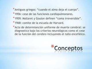
*
* Conciencia: representa la suma de las
actividades de la corteza cerebral.

* Despertar: estado de alerta o despierto mediada
por estructuras en tallo y talámicas mediales.

* Contenido: es la suma de las mentales cognitivas
y afectivas. Corteza y núcleos subcorticales
intactos

*
* Localizado en el puente, mesencéfalo, tálamo
e hipotálamo.

* La vía ascendente está formada por un grupo
de células monoaminérgicas en el tallo e
hipotálamo.

* El grupo de células colinérgicas están en el

pedúnculo, tegmento laterodorsal, núcleo
parabraquial que proyectan hacia el tálamo y
la corteza.

*
* Se caracteriza por la intensidad del estímulo que se
debe emplear para evocar una respuesta.

* Coma: no despierta, no responde, ojos cerrados.
* Estupor: dormido, responde a un fuerte estímulo, al
cesar, vuelve a dormir.

* Letargia y obnibulación: reducción de la alerta de
leve a moderada respectivamente.

*
* Coma: 2-4 semanas.
* Estado vegetativo persistente: > 1 mes.
* Neuropatologia:
* Traumático: lesión axonal difusa.
* No traumático: necrosis cortical laminar

*
* Estado de mínima conciencia: alteración severa
del estado de alerta, manifestaciones
cognitivas inconsistentes pero reproductibles.

* Mutismo akinético: inmóvil, ojos

cerrados, sueño-vigilia preservada, respuesta
nociceptiva mínima. Ausencia de signos que
comprometan las vías motoras.

* Locked-in síndrome:

cuadripleia, anartria, parálisis de la mirada
horizontal.
Table 3
Common Clinical Conditions Found in Patients in Coma With Their Clinical Findings
and Recommended Initial Management
Condition
Increased intracranial
pressure

Seizure activity

CNS infection

SAH

Liver failure

Recommended testing
and/or action

Possible findings
Papilledema, sixth cranial nerve
palsies, Kocher-Cushing
reflex (hypertension,
bradycardia, and irregular
respirations), presence of
vomitus
Eye deviation, pupillary
asymmetry, urinary or fecal
incontinence, tongue biting
Meningeal signs, fever,
petechial-purpuric rash
(meningococcemia), Osler’s
nodes (subacute bacterial
endocarditis), seizures, signs
of increased intracranial
pressure
Meningeal signs, third nerve
palsy or other cranial nerve
findings, subhyaloid
hemorrhages, papilledema or
other signs of increased
intracranial pressure
Icterus, ascites, gynecomastia,
spider nevi, decreased axillary
and pubic hair, hepatomegaly,
testicular atrophy, fetor
hepaticus

Imaging study
Depending on findings:
ventriculostomy placement,
intravenous mannitol,
hyperventilation
Immediate treatment with
antiepileptic drugs, followed
by EEG
Immediate initiation of
antibiotics, followed by
lumbar puncture

Head CT scanning
If CT equivocal, then lumbar
puncture

*

Liver function tests
including ammonia levels

CNS, central nervous system; EEG, electroencephalography; SAH, subarachnoid hemorrhage.
* Lesiones estructurales: masa hemisférica que

aumenta la PIC, herniación, compresión o
hemorragia en la parte superior del
mesencéfalo con involucro secundario del SARA

* Lesiones en tallo: daña SARA directamente.
* Lesiones que afectan ambos hemisferios y el
SARA

*
TABLE 51.3 Clinical Manifestations of Common Herniation Syndromes

Herniation Syndrome

Clinical Manifestatio ns

Central transtentorial

Impaired consciousness, abnormal respirations, symmetric small or midposition fixed or minimally reactive pupils,
decorticate evolving to decerebrat e posturing, rostrocaudal deterioration

Lateral transtentorial
(uncal)

Impaired consciousness, abnormal respirations, third nerve palsy (unilateral ly dilated pupil), hemiparesis ( may be false
localizing), rostrocaudal deterioration

Cerebellar tonsillar (foramen
magnum)

Impaired consciousness, neck rigidity, o pisthotonos, decerebrate rigidity, vo miting, irregular respirations, apnea,
bradycardia

Upward

Prominent brainstem signs, downward gaze deviat ion, upgaze palsy, decerebrate posturing (usu ally due to a
cerebellar mass lesion)

entral transtentorial herniation is due to symmetric downward displacement of the hemispheres causing
* Desórdenes convulsivos.
* Coma histérico: malingering, estados psicóticos
Table 4
Respiratory Patterns in Coma
Respiratory pattern

Typical underlying injury

Cheyne-Stokes

Waxing of frequency
followed by waning and
apnea

Central neurogenic
hyperventilation
Apneusis

Rapid and deep hyperpnea

Cluster breathing

Ataxic breathing

Kussmaul breathing

*

2–3 s long endinspiratory pauses,
sometimes alternating with
end-expiratory pauses
Periodic and irregular
frequency and amplitude of
respirations with irregular
pauses between clusters
Irregular and uneven
respirations with no
recognizable pattern
(preterminal states)
Deep, regular inspirations

Metabolic disturbances,
large bilateral hemispheric
insults, congestive heart
failure
Midbrain tegmentum
Midpons, caudal pons

Lower pontine and rostral
medullary lesions

Reticular formation of the
dorso-medial medulla

Metabolic acidosis

and decerebrate and as mentioned previously, posturing can also occur spontaneously. We recommend that practitioners describe the abnormal movements seen rather than just use the terms decorticate and decerebrate.
* Depende de 3 factores: causa, severidad y duración.
* No traumática: tóxica o metabólica >47% de
mortalidad  estado vegetativo persistente

* EVC >70% de mortalidad.
* Traumático: >49% de mortalidad.
* 70% tienen reintegración social; 20% dependientes.

*
* TBI: GCS 70% del VPP para pronóstico.

* Hallazgos motores: si en 6 horas no mejoran, la

mortalidad es del 63% (flexor, 83% (extensión o
flacidez).
* Edad: mas severo con la edad.
* Signos neuro oftálmicos: >70% VPP; 95% 6hr.
* Daño secundario: hipotensión, hipoxia, PIC dobla
mortalidad
* Imagen: TC ver lesiones focale o daño difuso;
70% VPP
.
* Duración del coma.
* EEG y SSEPs.
* Marcadores biológicos: proteína ácida fibrilar
glial, esqueleto astroglial y S100B.
*
*
* Antiguos griegos: “cuando el alma deja el cuerpo”.
* 1956: cese de las funciones cardiopulmonares.
* 1959: Mollaret y Goulon definen “coma irreversible”.
* 1968: comite de la escuela de Harvard.
* Acta de determianción uniforme de muerte cerebral: se

diagnostica bajo los criterios neurológicos como el cese
de la función del cerebro incluyendo el tallo encefálico.

*
* Adultos:

* TCE.
* Hemorragia subracnoidea.
* Daños hipóxico-isquémico.
* Falla hepática fulminante.

* Niños:

* Abuso.
* Accidentes en vehículos de motor.
* Asfixia.

*
* Evidencia clínica o por neuroimagen de catástofre en
SNC que correlacione MC.

* Complicaciones médicas.
* Sin evidencia de intoxicación, bloqueo neuromuscular
* Temperatura > 32ºC.
* Hipotensión significativa ausente.

*
* Determinar estado de coma.
* Signo de Lázaro.
* Reflejos de tallo: pupilas de 4? y 9 mm, pruebas
calóricas.
* Prueba de la apnea.

* Considerar:
* Trauma facial.
* Alteraciones pupilares
* previas.
* Niveles tóxicos.
* SAOS: PCO2 elevado.

*
*
312

Georg

Table 6
The Use of Transcranial Doppler for Brain Death Confirmation
Subjects:
Patients who fulfill clinical criteria for diagnosis of brain death and do not have ventricular
drains or large craniotomies that can prevent increases in intracranial pressure.
Examination technique:
Minimal requirement: Bilateral insonation with assessment of at least one artery on each side.
Recommended: Bilateral intra- and extracranial examination, performed twice with a 30-min
interval. Extracranial examination should include the internal and common carotid arteries
and the vertebral arteries.
Intracranial flow patterns consistent with brain death:
Reverberating (oscillating) flow or
Low amplitude (<50 cm/s), brief (<200 ms) spikes in early systole without diastolic flow or
absent intracranial flow in a patient who is known to have a temporal window. It is recommended to further confirm the diagnosis with a study of the extracranial vessels.
* Potenciales somatosensoriales: ausencia

bilateral de N20-P22 a la estimulación del
nervio mediano.
* Definir clinicamente el estado de alerta y
conciencia.

* Identificar las causas subyacentes y las lesiones
secundarias.

* Realizar los algoritmos descritos en las guías.

*

Coma y muerte cerebral

  • 1.
  • 2.
    * Conciencia: representala suma de las actividades de la corteza cerebral. * Despertar: estado de alerta o despierto mediada por estructuras en tallo y talámicas mediales. * Contenido: es la suma de las mentales cognitivas y afectivas. Corteza y núcleos subcorticales intactos *
  • 3.
    * Localizado enel puente, mesencéfalo, tálamo e hipotálamo. * La vía ascendente está formada por un grupo de células monoaminérgicas en el tallo e hipotálamo. * El grupo de células colinérgicas están en el pedúnculo, tegmento laterodorsal, núcleo parabraquial que proyectan hacia el tálamo y la corteza. *
  • 4.
    * Se caracterizapor la intensidad del estímulo que se debe emplear para evocar una respuesta. * Coma: no despierta, no responde, ojos cerrados. * Estupor: dormido, responde a un fuerte estímulo, al cesar, vuelve a dormir. * Letargia y obnibulación: reducción de la alerta de leve a moderada respectivamente. *
  • 5.
    * Coma: 2-4semanas. * Estado vegetativo persistente: > 1 mes. * Neuropatologia: * Traumático: lesión axonal difusa. * No traumático: necrosis cortical laminar *
  • 6.
    * Estado demínima conciencia: alteración severa del estado de alerta, manifestaciones cognitivas inconsistentes pero reproductibles. * Mutismo akinético: inmóvil, ojos cerrados, sueño-vigilia preservada, respuesta nociceptiva mínima. Ausencia de signos que comprometan las vías motoras. * Locked-in síndrome: cuadripleia, anartria, parálisis de la mirada horizontal.
  • 8.
    Table 3 Common ClinicalConditions Found in Patients in Coma With Their Clinical Findings and Recommended Initial Management Condition Increased intracranial pressure Seizure activity CNS infection SAH Liver failure Recommended testing and/or action Possible findings Papilledema, sixth cranial nerve palsies, Kocher-Cushing reflex (hypertension, bradycardia, and irregular respirations), presence of vomitus Eye deviation, pupillary asymmetry, urinary or fecal incontinence, tongue biting Meningeal signs, fever, petechial-purpuric rash (meningococcemia), Osler’s nodes (subacute bacterial endocarditis), seizures, signs of increased intracranial pressure Meningeal signs, third nerve palsy or other cranial nerve findings, subhyaloid hemorrhages, papilledema or other signs of increased intracranial pressure Icterus, ascites, gynecomastia, spider nevi, decreased axillary and pubic hair, hepatomegaly, testicular atrophy, fetor hepaticus Imaging study Depending on findings: ventriculostomy placement, intravenous mannitol, hyperventilation Immediate treatment with antiepileptic drugs, followed by EEG Immediate initiation of antibiotics, followed by lumbar puncture Head CT scanning If CT equivocal, then lumbar puncture * Liver function tests including ammonia levels CNS, central nervous system; EEG, electroencephalography; SAH, subarachnoid hemorrhage.
  • 9.
    * Lesiones estructurales:masa hemisférica que aumenta la PIC, herniación, compresión o hemorragia en la parte superior del mesencéfalo con involucro secundario del SARA * Lesiones en tallo: daña SARA directamente. * Lesiones que afectan ambos hemisferios y el SARA *
  • 10.
    TABLE 51.3 ClinicalManifestations of Common Herniation Syndromes Herniation Syndrome Clinical Manifestatio ns Central transtentorial Impaired consciousness, abnormal respirations, symmetric small or midposition fixed or minimally reactive pupils, decorticate evolving to decerebrat e posturing, rostrocaudal deterioration Lateral transtentorial (uncal) Impaired consciousness, abnormal respirations, third nerve palsy (unilateral ly dilated pupil), hemiparesis ( may be false localizing), rostrocaudal deterioration Cerebellar tonsillar (foramen magnum) Impaired consciousness, neck rigidity, o pisthotonos, decerebrate rigidity, vo miting, irregular respirations, apnea, bradycardia Upward Prominent brainstem signs, downward gaze deviat ion, upgaze palsy, decerebrate posturing (usu ally due to a cerebellar mass lesion) entral transtentorial herniation is due to symmetric downward displacement of the hemispheres causing
  • 11.
    * Desórdenes convulsivos. *Coma histérico: malingering, estados psicóticos
  • 12.
    Table 4 Respiratory Patternsin Coma Respiratory pattern Typical underlying injury Cheyne-Stokes Waxing of frequency followed by waning and apnea Central neurogenic hyperventilation Apneusis Rapid and deep hyperpnea Cluster breathing Ataxic breathing Kussmaul breathing * 2–3 s long endinspiratory pauses, sometimes alternating with end-expiratory pauses Periodic and irregular frequency and amplitude of respirations with irregular pauses between clusters Irregular and uneven respirations with no recognizable pattern (preterminal states) Deep, regular inspirations Metabolic disturbances, large bilateral hemispheric insults, congestive heart failure Midbrain tegmentum Midpons, caudal pons Lower pontine and rostral medullary lesions Reticular formation of the dorso-medial medulla Metabolic acidosis and decerebrate and as mentioned previously, posturing can also occur spontaneously. We recommend that practitioners describe the abnormal movements seen rather than just use the terms decorticate and decerebrate.
  • 15.
    * Depende de3 factores: causa, severidad y duración. * No traumática: tóxica o metabólica >47% de mortalidad  estado vegetativo persistente * EVC >70% de mortalidad. * Traumático: >49% de mortalidad. * 70% tienen reintegración social; 20% dependientes. *
  • 16.
    * TBI: GCS70% del VPP para pronóstico. * Hallazgos motores: si en 6 horas no mejoran, la mortalidad es del 63% (flexor, 83% (extensión o flacidez). * Edad: mas severo con la edad. * Signos neuro oftálmicos: >70% VPP; 95% 6hr. * Daño secundario: hipotensión, hipoxia, PIC dobla mortalidad * Imagen: TC ver lesiones focale o daño difuso; 70% VPP . * Duración del coma. * EEG y SSEPs. * Marcadores biológicos: proteína ácida fibrilar glial, esqueleto astroglial y S100B.
  • 17.
  • 25.
  • 26.
    * Antiguos griegos:“cuando el alma deja el cuerpo”. * 1956: cese de las funciones cardiopulmonares. * 1959: Mollaret y Goulon definen “coma irreversible”. * 1968: comite de la escuela de Harvard. * Acta de determianción uniforme de muerte cerebral: se diagnostica bajo los criterios neurológicos como el cese de la función del cerebro incluyendo el tallo encefálico. *
  • 27.
    * Adultos: * TCE. *Hemorragia subracnoidea. * Daños hipóxico-isquémico. * Falla hepática fulminante. * Niños: * Abuso. * Accidentes en vehículos de motor. * Asfixia. *
  • 28.
    * Evidencia clínicao por neuroimagen de catástofre en SNC que correlacione MC. * Complicaciones médicas. * Sin evidencia de intoxicación, bloqueo neuromuscular * Temperatura > 32ºC. * Hipotensión significativa ausente. *
  • 29.
    * Determinar estadode coma. * Signo de Lázaro. * Reflejos de tallo: pupilas de 4? y 9 mm, pruebas calóricas. * Prueba de la apnea. * Considerar: * Trauma facial. * Alteraciones pupilares * previas. * Niveles tóxicos. * SAOS: PCO2 elevado. *
  • 31.
  • 35.
    312 Georg Table 6 The Useof Transcranial Doppler for Brain Death Confirmation Subjects: Patients who fulfill clinical criteria for diagnosis of brain death and do not have ventricular drains or large craniotomies that can prevent increases in intracranial pressure. Examination technique: Minimal requirement: Bilateral insonation with assessment of at least one artery on each side. Recommended: Bilateral intra- and extracranial examination, performed twice with a 30-min interval. Extracranial examination should include the internal and common carotid arteries and the vertebral arteries. Intracranial flow patterns consistent with brain death: Reverberating (oscillating) flow or Low amplitude (<50 cm/s), brief (<200 ms) spikes in early systole without diastolic flow or absent intracranial flow in a patient who is known to have a temporal window. It is recommended to further confirm the diagnosis with a study of the extracranial vessels.
  • 38.
    * Potenciales somatosensoriales:ausencia bilateral de N20-P22 a la estimulación del nervio mediano.
  • 39.
    * Definir clinicamenteel estado de alerta y conciencia. * Identificar las causas subyacentes y las lesiones secundarias. * Realizar los algoritmos descritos en las guías. *

Notas del editor

  • #3 Escaracterizadapor la conciencia de simismo y sumedioambiente y la habilidad de responder a el y a estimulosintrinsecos
  • #4 Impairment of consciousness is caused by damage to those structures or by bilateral hemispheric damage
  • #5 Ambos terminos son ambiguos
  • #6 Estado de despierto sin funcionescognitivas In a recent study, Adams et al. proposed that the fundamental structural abnormality underlying the persistent vegetative state, regardless of etiology, is widespread damage to subcortical white matter and the thalami .
  • #7 Ausencia de rigidez y espasticidad, puedeparpedearDestruccion del puene ventralInfarto, homorragia, mielinolisisMutismovssindrome lento? Mutismopor CairnsAnartria: (Del griego an-, privación y arthron, articulación). (Pierre Marie). Sinónimo: AFASIA MOTRIZ SUBCORTICAL (Déjerine). Trastorno del lenguajequeconsisteúnicamente en la imposibilidad de articular los sonidos. El pacienteafecto de anartriacomprende lo que se le dice, lo puede leer, pero no puedepronunciar la palabraque lee; puedeescribir, puedetambiénindicarporpresiones de mano o porcualquierotrosigno el número de sílabascontenidas en la palabraque no puedepronunciar. La anartriaobedece a unalesión cerebral focal localizada en la zona del núcleo lenticular.
  • #9 Descartaralteracionesmetabólicas, tóxicas.Estudios de imagen, tratarsiesposible.Darglucosa o tiaminaIsqueiatrombolizar, hemorragiacerebelo o epiduralA “coma cocktail,” consisting of dextrose, flumazenil, naloxone, and thiamine is sometimes used in the initial management of the comatose
  • #11 During rostrocaudal deterioration, neurologic dysfunction becomes progressively more dramatic. Clinical stages occur as if the brain had been transversely sectioned at a particular level (diencephalon, midbrain, pons, or medulla)
  • #12 In addition to alteration of consciousness during the ictus, seizure disorders may also cause AMS due to postictal unresponsiveness, absence status, psychomotor status, and subclinical status epilepticus. In the postictal period there is often depression of consciousness, a desire to sleep, confusion, and disorientation
  • #16 The absence of brainstem reflexes, a low Glasgow Coma Scale score, and the presence of hypoxia or hypotension during the first 24 h after admission are further very important prognostic factors. La severidad del coma por el glasgowes factor predictivo a las 6 hrs de establecimiento. Cuandoes el tce de leve a moderado, caue el 10% en la admision y 7% al 3er dia y 2% al dia 14.
  • #17 Ausencia de sxocularesmurieron a las 6 hrs. ref 18Ssep se confunden con la sedacion, 100% especificidad en la ausencia cortical bilateralP300 al decirpalabrascomomomiacomo predictor en GOS 3, no ha diferencia con otrosestudios en pxquerecobraronlasfuncionessuperiores.Proteina glial como predictor de mortalidadOtroestudio: mediciones a los 7 dias de la relacion de glutamoto/glutamina y colinafuesignificativamenteelevada en la sustanciablanca occipital y parietal de 6-12 meses de outcome con GOS menor a 4
  • #23 Figure 9–5. Akineticmutism seen in Patient 9–2. (A) Computed tomography scan demonstrating large mesencephalic mass with surrounding edema. (B) Series of magnetic resonance axial images following treatment with steroids and reduction of mesencephalic lesion. Middle image shows high-signal abnormalities in the ventral midbrain. (C) Single photon emission tomography imaging demonstrates diffuse cerebral hypoperfusion with relative sparing of cerebellar blood flow.
  • #27 1959: Presentaron 23 pxdiscussed hallmarks of brain death such as the absence of brainstem function, the absence of spontaneous respiration, the cardiovascular collapse that ensued without the use of pressors and many of the common associated medical complications.
  • #29 Estanderes medicos aceptadosporQuality Standards Subcommittee of the American Academy of Neurology in 1995 en mayores de 18 años- Edema difuso, masas o herniacion, hacer PLAlteraciones en ES o endocirnas
  • #30 Respiratory-like movements, such as shoulder elevation and adduction, back arching, and intercostal expansion, without significant tidal volumes can also be acceptedThe eyes must be observed for 1–3 min after irrigation and 5 min must be allowed between testing on each side
  • #34 Eeg: 0.3-0.4 comoconstante de tiempo, ekg o electrodo en el dorso de la manoparadetectaractividadextracerebral, repetirsi hay silencios, no portelefono
  • #37 As intracranial pressure (ICP) rises, it reaches values above the end-diastolic arterial blood pressure (ABP) and therefore starts compromising diastolic flow (middle portion, top figure). When ICP exceeds the diastolic ABP, diastolic flow ceases and only systolic peaks are seen (right end, top figure). Subsequently there is reverberating (oscillating) flow. The biphasic flow velocity spectrum consists of equivalent, opposing inflow and outflow components and the resulting time-averaged mean velocity is zero, i.e., effectively there is already circulatory arrest. In final stages flow consists only of very small brief spikes in early systole followed by no flow at all. The angiographic correlate of oscillating flow and systolic spikes is usually stasis filling (delayed and tapered filling of the intracranial arteries without venous drainage after 26 s). When Doppler signals are absent, angiography usually shows no filling of the intracranial vessels