Epistaxis rinologia
•Descargar como PPTX, PDF•
3 recomendaciones•837 vistas

Epistaxis, modulo rinologia Cummings

Recomendados
Más contenido relacionado
La actualidad más candente
La actualidad más candente (20)
Destacado
Destacado (16)
Similar a Epistaxis rinologia
Similar a Epistaxis rinologia (20)
Más de Angel Castro Urquizo
Más de Angel Castro Urquizo (20)
Último
Último (20)
Epistaxis rinologia
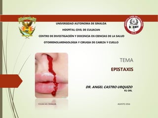
- 1. TEMA EPISTAXIS UNIVERSIDAD AUTONOMA DE SINALOA HOSPITAL CIVIL DE CULIACAN CENTRO DE INVESTIGACIÓN Y DOCENCIA EN CIENCIAS DE LA SALUD OTORRINOLARINGOLOGIA Y CIRUGIA DE CABEZA Y CUELLO DR. ANGEL CASTRO URQUIZO R1 ORL CULIACAN SINALOA AGOSTO 2016
- 2. Introducción EPI = Sobre, encima STAZO = Fluir Gota a Gota Urgencia ORL mas común Afecta al 60% de la población en su vida 10% atención medica Hombres < 10 años - > 40 años Invierno .
- 4. Anatomía • 1.- A. Facial A. Labial superior • 2.- A. Maxilar Interna A. Esfenopalatina A. Palatina descendente A. Faringea A. Infraorbitaria A. Alveolar superior A. Vidiana Carótida externa • 1.- Oftálmica Etmoidales Carótida Interna Septum anterior
- 5. Anatomía vascular Sistema arterial de la carótida externa Facial - Labial superior - Angular Maxilar Interna -Esfenopalatina -Conchal (Posterolatera) -Septal (Posteromedial) -Palatina Descendente -Palatina mayor (Septum Anteroinferior)
- 6. Anatomía vascular Sistema arterial de la carótida interna 1.- Oftálmica -Etmoidales -Anterior (septum anterosuperior) -Posterior (septum posterosuperior, pared lateral nasal)
- 8. Anatomía vascular Plexos Plexo Keisselbach´s • Esfenopalatina • Palatina mayor • Labial superior • Etmoidal anterior
- 11. Clasificación topográfica Anterior • 90% • Área anterior del tabique Superior • Ramas las A. etmoidal anterior o posterior • Fracturas nasales Posterior • A. Esfenopalatina • Difíciles de controlar todo BIEN Dagueado
- 13. Etiologia Primarias • Idiopáticas Secundarias • Locales • Sistémicas
- 14. Etiología Causas secundarias Locales • Trauma • Cirugías • Deformidades anatómicas • Tumores • Cuerpo extraño • Causas Inflamatorias • Causas granulomatosas Sistémicas • Drogas y fármacos • Enfermedades hematológicas • HAS • Enfermedades hepatobiliares • Alcoholismo • Enfermedades tejido conectivo/ vascular • Malnutrición
- 16. • Rinosinusitis • Rinitis Seca • Rinitis Alérgica • Rinitis Vasomotora • Rinofaringitis Inflamatorias • Viral • Bacteriana • Micótica • Parasitosis Rinitis Infecciosas • Rascado Nasal(1) • Fx Nasales • Fx del Piso Craneal • Qx, Iatrogénicos • Cuerpos extraños Traumatismos
- 17. Granulomatosas Wegener TB Sarcoidosis
- 18. • Desviación Septal • Desviación Rinoseptal • Perforación Septal Estructurales • Pólipos • Papiloma Invertido • Angiofibroma, Hemangiomas, ADC • Rabdomiosarcoma, Linfagiomas Neoplasias • Medicamentos locales vasoconstrictores (oximetazolina) • Drogas inhaladas (cocaína, tabaco, marihuana) • Telangiectasia Hemorrágica Hereditaria Vasculares
- 25. Etiología Causas secundarias sistémicas • Deficiencia del factor VIII • Deficiencia del factor IX Coagulopatías • Leucemias • Linfomas • Anemia Aplásica Neoplasias • HTAVasculares Otros Diabetes Mellitus Aspirina, Warfarina, etcMedicamentos Sistémicos
- 27. Evaluación clínica Anamnesis: Intensidad Anterior o Posterior Nueva o recurrente Espontánea o provocada. Examen Físico: Signos Vitales Localizar el área de sangrado Laboratorio: HC, Plaquetas, TP y TPT. Orientar sobre causas, pronóstico y complicaciones
- 29. Manejo inicial Calcular perdidas SV Aspecto físico Umbral 9g/dl Laboratoriales
- 30. Manejo Clínico Vía aérea • Volumen y soporte hemodinámico Posición • 15-20 min. Vasoconstrictor tópico
- 32. Examinación directa Tratar establecer el sitio Nitrato de plata, Bipolar Trabajar protegidos Si no se logra localizar endoscopio
- 33. Manejo clínico
- 34. Collagen-derived particles con trombina bovina
- 35. Manejo clínico No sonarse 2 semanas Cremas Anestesia Cauterización endoscópica
- 36. Epistaxis idiopática posterior Septum (Ramas septal esfenopalatina) Meato medio e inferior
- 37. Manejo clínico Taponamiento anterior Gasa serpentina + cloranfenicol ungüento Adecuada anestesia y vasoconstricción tópica. Tapón se deja entre 5 y 7 días. Antibióticos de amplio espectro.
- 40. Manejo clínico Taponamiento Posterior No es controlada eficazmente por taponamiento anterior. Sangrado profuso Formas: – Sonda de Foley – Tapón con paquete de gasa Posterior epistaxis tasa de fallo 26% and 52% Tasa de complicaciones: 2% and 68.8%.
- 41. Manejo clínico Taponamiento Posterior Sonda de Foley Sonda de Foley No.14 hasta que su extremo se vea por detrás de la úvula. Se infla el balón con 10 ml de agua y se tracciona suavemente hasta que el globo quede anclado a las coanas
- 44. Manejo clínico Complicaciones del manejo Perforación septal Necrosis Apnea, Hipoxia Shock hipovolémico Shock toxico staphylococcico Daño neurovascular Celulitis periorbitaria Rinosinusitis Miasis
- 47. Manejo quirúrgico Ligaduras vasculares Ligadura maxilar • Éxito 90% • Cadwell-Lucfosa pterigomaxilar Ligadura carótida externa • Severo • Compromiso vital • EVC
- 48. Manejo quirúrgico Ligaduras vasculares Ligadura Esfenopalatina endoscópica • Menos morbilidad que en resto • Preferida en posterior • Disección posterior cornete medio • 92% éxito
- 49. Ligadura Esfenopalatina 97% - 2 o mas ramas mediales a la cresta etmoidal 67% 3 o mas ramas 35% 4 o mas ramas
- 50. * *
- 51. Ligadura Esfenopalatina Complicaciones Persistencia de epistaxis Costras nasales Entumecimiento del paladar RSN Disminución de lagrimeo Perforación septal
- 52. Manejo quirúrgico Embolización Maxilar esfenopalatina 71-95% control 27% Complicaciones EVC Parálisis facial Ceguera Muerte
- 54. Sangrado de arteria etmoidal anterior Fx nasoetmoidales -Sangrado intraorbitario -Intracraneal Dehiscencia 11-40% 1-2mm detrás de la unión de la pared posterior del receso frontal y celdillas etmoidales ant.
- 55. Irrigación con agua caliente 40-46 C° sin cambios histologicos 46-52 C° edema de la mucosa > 52 C° Necrosis 500 ml 3 minutos Paciente sentado Éxito 82%
- 56. Manejo de epistaxis en pacientes con HHT Terapia hormonal anticuerpo monoclonal contra VEGF-A Antifibrinoliticos Coagulación laser Septodermoplastia
Notas del editor
- CM 42
- Afecta al 60% y 10% requieren asistencia medica Distribución bimodal Urgencias que amenaz la vida
- Car. Ext. … tiroidea sup, lingual, facial, occipital, auricular posterior, faríngea inf.
- Estan en el mucoperiostio y pericondrio VIDIANA - GRAN ANASTOMOSIS ENTRE Carotiad interna y esfenopalatina
- Labial superior columnell,a pared laeteral, septum anterior, Angular punta y dorso MI(fosa pterigopalatina)Esfenopalatina pared lateral, septum posterior Palatina descendente palatina menor Palatina mayor: septum,
- 1.- Oftalmica (fisura supraorbitaria).- Etmoidales ant y post(techo etmoidal) ANTERIOR porción lateral y 1/3 anterior POSSTErIOR cornete superior, septum posterior
- Endoscopic view of the left skull base in a latex-injected specimen, showing the course of the anterior ethmoid artery. A, Zürich microscissors divide the artery close to the orbit. B, Endoscopic view of the left skull base area after a median drainage procedure shows the anterior nasal branch coming off the ethmoid artery and the first olfactory fiber just behind (arrows
- Kieselbach: Ramas esfenopalatina, palatina mayor, labial superior, ramas etmoidal anterior Plezo woodroof. Plexo nasofaríngeo: Etmoidea posterior, Nasal posterior, SPA, Faringea ascendente
- Linea imaginariaEntre el ostiom
- Idiopaticas mayoria
- La cauma mas común, manipulación, trauma digital Cirugia: sinus, endoscópica, base cráneo Deformidades: desviación septal Tumores: bbenignos, vasculares, malignos Drogas: aines, ASA,
- Endoscopic view (A) and computed tomography scan (B) of a cavernous hemangioma arising from the olfactory cleft on the right side that has caused a severe epistaxis.
- 1.- traumatico autoinducido 2.- medicamentos topicos 3.- inhalacion cocaina
- Endoscopic view of Wegener granulomatosis. po de vasculitis poco frecuente en la cual se produce hinchazón e inflamación de losvasos sanguíneos, dificultando así el flujo de la sangre. Afecta principalmente los vasos sanguíneos en lanariz, senos paranasales, oídos, pulmones y riñones,
- Angiofibroma nasofaríngeo juvenil: obstrucción y epistaxis unilateral Adc. adenocarcinoma
- Granuloma pyogenoPyogenic granuloma on the septum, right side nasal vestibule.
- Endoscopic view of an angiofibroma occluding the right nasal cavity and choana.
- Squamous cell carcinoma of the right nasal cavity originat
- Total septal perforation causing epistaxis and crusting in a patient who had abused cocaine.
- Telangiectasia Hemorrágica Hereditaria AD
- Hemofilia A VIII, B IX
- TRANQUILIZAR Importanet. 1ero signos vitales.. Controlar la TA
- TA signo tardio
- Vitamin K takes more than 6 hours to work, and it can delay anticoagulation for 7 days after warfarin is started. If the international normalized ratio is greater than 4, the warfarin should be stopped, and fresh frozen plasma should be given.3
- NASAL ENDOSCOPY Nasal endoscopy has a vital role in defining and treating posterior epistaxis, and it can substantially reduce hospital admissions.17-19
- Solo en sangrados anteriores The majority of posterior idiopathic bleeds are from the septum, usually from the septal branch of the sphenopalatine artery as it runs submucosally
- Caja con
- PREVIA FENILEFRINA Y COCAINA (DESCONGESTION EN 5 MIN Y ANESTESIA EN 9 MIN) AYUDA MUCHO No recomendado en bilateral Cremas antisépticas por 2 semanas
- LUGARES EN EPISTAXIS POSTERIOR Endoscopic view of the bleeding site in severe epistaxis coming from a conchal branch of the sphenopalatine artery on the right side, just before monopolar suction cautery was performed.
- CUANDO Electrocauterio no es efectivo o no se localiza el sitio de sangrado El tapón se introduce formando pliegues que semejan el fuelle de un acordeón. 2. Se empieza por el piso de la fosas nasales y se termina en el ático. 3. Porciones anteriores de las cámaras nasales, hasta las coanas si es necesario. (dos puntas por fuera de las narinas). 4. Debe quedar suficientemente apretado para cohibir el sangrado.
- STS > 24 horas, exotoxina
- Necrosis of the columella after bilateral posterior nasal packing. One study concluded that systemic prophylactic antibiotics are unnecessary in the majority of epistaxis patients who have nasal packs.61 The use of topical antibiotics may be more appropriate, cheaper, and just as effective.
- Cara anterior del seno maxilar y después la fosa pterigomaxilar ENDOSCOPICA LIGADURA CAROTIDA– EN OCASIONES INEFECTIVA POR ANOSTOMOSIS Y X LA IRRIGACION CONTRALATERAL
- If bleeding cannot be controlled after endoscopic examination and cautery and/or nasal packing, sedation and examination under a local or general anesthetic are indicated. Bipolar diathermy of any bleeding points or ESPAL is then the treatment of choice.10
- Importante tomar en consideración que entra ala nariz en varias ramas Los fallos se dan por ligar o cauterizar solo una rama
- ndoscopic view of the right ethmoid crest (asterisk) in a human skull. B, View of the right lateral wall of the nose injected with pink latex to show four branches of the sphenopalatine artery, three above and one below the ethmoid crest (asterisk). Intraoperative endoscopic view of the dissection of the sphenopalatine artery just behind the ethmoid crest (asterisk). Bipolar cautery of the first branch is performed just below the ethmoid crest. B, Final view after cautery of all four branches, two below and two above the ethmoid crest, and suction within the right sphenoid sinus.
- We do not advocate packing and repacking for refractory epistaxis if this can be avoided
- EFECTIVA CAR. EXT. PELIGROSA EN CAR. INT
- Bilateral superselective angiograms of the sphenopalatine arteries. A, The crossover anastomosis of the septal mucosa artery supply. B, Postembolization view.
- Asociado con fx nasoetmoidal The bony canal in which the anterior ethmoid artery crosses the skull base can be dehiscent, making it vulnerable to damage.90 A dehiscence of the anterior ethmoid artery of 11% to 40% has been reported.91 Intraoperative endoscopic view of bipolar suction cautery of the left anterior ethmoid artery in epistaxis. Partial anterior ethmoidectomy and exposure of the vessel just below the skull base is shown.
- Se introdujo en sangrados pospartos EDEMA --- COMPRESION DE VASOS Y PROPAGA CASCADA COAGULACION Éxito sin complicacsiones: 82%
- Hereditary hemorrhagic telangiectasia (HHT) is an autosomal-dominant multisystemic disorder characterized by telangiectasia that affects cutaneous and mucosal surfaces and arteriovenous malformations in the pulmonary, cerebral, and hepatic circulations . The most common symptom in this population is epistaxis, which affects more tha 90% of individuals. noretindrona/estradiol y danazo Bevacizumab que consiste en la eliminación de la mucosa nasal original y su sustitución por un injerto de piel que normalmente se toma del muslo y a veces de la mucosa nasal.