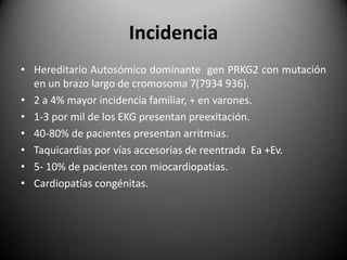
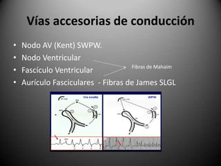
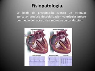
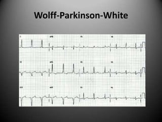
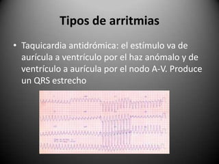
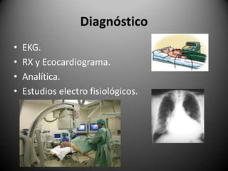
Este documento resume la enfermedad de Wolff-Parkinson-White, una afección cardíaca hereditaria caracterizada por la presencia de vías accesorias de conducción que causan una conducción anómala entre las aurículas y los ventrículos. Se detallan la incidencia, las vías accesorias más comunes, los síntomas, los hallazgos en el electrocardiograma, los tipos de arritmias asociadas y los enfoques de diagnóstico y tratamiento, incluida la ablación por catéter de radiofrecuencia
Diagnóstico Electrocardiográfico de Infarto de Miocardio en presencia de Bloq...Ernest Spitzer
Revisión breve respecto a la utilidad del electrocardiograma en el diagnóstico de Infarto de Miocardio en presencia de Bloqueo de Rama Izquierda y Marcapasos. Se describen los beneficios y limitaciones de los criterios de Sgarbossa, Wackers y otros.
Early repolarization (ER), consisting of a J-point elevation, notching or slurring of the terminal portion of the R wave (J wave), and tall/symmetric T wave, is a common finding on the 12-lead electrocardiogram. For decades, it has been considered as benign, barring sporadic case reports and basic electrophysiology research that suggested a critical role of the J wave in the pathogenesis of idiopathic ventricular fibrillation (VF). In 2007-2008, a high prevalence of ER in patients with idiopathic VF was reported and subsequent studies reinforced the results. This PPT describes the current state of knowledge concerning ER syndrome associated with sudden cardiac death.
Aula sobre a Síndrome de Wolff Parkinson-White , ministrada no I Simpósio Catarinense de Arritmia Cardíaca, realizado em Julho de 2017, em Florianópolis - SC.
O evento, promovido pela Clínica Ritmo, clínica especializada no tratamento de Arritmias e Implante de Marcapasso, teve como objetivo abordar todas as formas de arritmias cardíacas e as possibilidades de tratamentos, com temas trazidos a partir de casos reais tratados pelos especialistas da Clínica Ritmo nos últimos cinco anos.
Para saber mais sobre os procedimentos, acesse: http://www.clinicaritmo.com.br/
Tachycardias are broadly categorized based upon the width of the QRS complex on the electrocardiogram (ECG). A narrow QRS complex (<120 milliseconds) reflects rapid activation of the ventricles via the normal His-Purkinje system, which in turn suggests that the arrhythmia originates above or within the His bundle (ie, a supraventricular tachycardia). The site of origin may be in the sinus node, the atria, the atrioventricular (AV) node, the His bundle, or some combination of these sites. A widened QRS (≥120 milliseconds) occurs when ventricular activation is abnormally slow. The most common reason that a QRS is widened is because the arrhythmia originates below the His bundle in the bundle branches, Purkinje fibers, or ventricular myocardium (eg, ventricular tachycardia). Alternatively, a supraventricular arrhythmia can produce a widened QRS if there are either pre-existing or rate-related abnormalities within the His-Purkinje system (eg, supraventricular tachycardia with aberrancy), or if conduction occurs over an accessory pathway. Thus, wide QRS complex tachycardias may be either supraventricular or ventricular in origin.
Wolff–Parkinson–White syndrome (WPW) is one of several disorders of the conduction system of the heart that are commonly referred to as pre-excitation syndromes. WPW is caused by the presence of an abnormal accessory electrical conduction pathway between the atria and the ventricles. Electrical signals travelling down this abnormal pathway (known as the bundle of Kent) may stimulate the ventricles to contract prematurely, resulting in a unique type of supraventricular tachycardia referred to as an atrioventricular reciprocating tachycardia.The incidence of WPW is between 0.1% and 0.3% in the general population.Sudden cardiac death in people with WPW is rare (incidence of less than 0.6%), and is usually caused by the propagation of an atrial tachydysrhythmia (rapid and abnormal heart rate) to the ventricles by the abnormal accessory pathway.
Wolff–Parkinson–White syndrome (WPW) is one of several disorders of the conduction system of the heart that are commonly referred to as pre-excitation syndromes. WPW is caused by the presence of an abnormal accessory electrical conduction pathway between the atria and the ventricles. Electrical signals traveling down this abnormal pathway (known as the bundle of Kent) may stimulate the ventricles to contract prematurely, resulting in a unique type of supra-ventricular tachycardia referred to as an atrio-ventricular reciprocating tachycardia.
se aborda el concepto de respiración, sus fases, su fisiopatología, hipoventilación, clasificación, manifestaciones clínicas, tratamiento, oxigenoterapia.
Definición, Etiologia, Patogenia, Factores de riesgo, Manifestaciones y Diagnóstico basados en ECG y Tratamiento de los diferentes tipos de taquiarritmias Basado en la Medicina Interna De Harrison 19va edición
STEMI es el acrónimo de “ST elevation myocardial infartion” y hace referencia a lo que en español conocemos como SCACEST “Síndrome coronario agudo con elevación del ST”. Por su parte el término NSTEMI hace referencia a lo que conocemos con SCASEST, “Síndrome coronario agudo sin elevación del ST” ó “No ST elevation myocardial infartion”.
La elevación del segmento ST se traduce como la lesión subepicárdica o transmural acontecida por isquemia mantenida que provoca un retraso en la repolarización de todo el miocardio de la región afectada generando en el EKG esta alteración. El ascenso del ST en un EKG nos pone en alerta ante un infarto de miocardio con oclusión completa que requiere de una revascularización urgente.
No obstante, bajo este paradigma diagnóstico del STEMI actual (valorando únicamente ascensos del ST), encontramos que el 25-30% de los pacientes NSTEMI sí que tienen oclusión total en el cateterismo cardíaco tardío. Encontramos además, que si en vez de usar criterios diagnósticos de STEMI estrictos, se realiza una interpretación experta del ECG, los cardiólogos pueden reclasificar con éxito al 28 % de los pacientes con NSTEMI como pacientes con oclusión coronaria aguda que responden a la terapia de reperfusión inmediata, lo que reduce a la mitad la mortalidad a corto y largo plazo.
REALIZAR EL ACOMPAÑAMIENTO TECNICO A LA MODERNIZACIÓN DEL SISCOSSR, ENTREGA DEL SISTEMA AL MINISTERIO DE SALUD Y PROTECCIÓN SOCIAL PARA SU ADOPCIÓN NACIONAL Y ADMINISTRACIÓN DEL APLICATIVO, EN EL MARCO DEL ACUERDO DE SUBVENCIÓN NO. COL-H-ENTERRITORIO 3042 SUSCRITO CON EL FONDO MUNDIAL.
DIFERENCIAS ENTRE POSESIÓN DEMONÍACA Y ENFERMEDAD PSIQUIÁTRICA.pdfsantoevangeliodehoyp
Libro del Padre César Augusto Calderón Caicedo sacerdote Exorcista colombiano. Donde explica y comparte sus experiencias como especialista en posesiones y demologia.
2. Incidencia Hereditario Autosómico dominante gen PRKG2 con mutación en un brazo largo de cromosoma 7(7934 936). 2 a 4% mayor incidencia familiar, + en varones. 1-3 por mil de los EKG presentan preexitación. 40-80% de pacientes presentan arritmias. Taquicardias por vías accesorias de reentrada Ea +Ev. 5- 10% de pacientes con miocardiopatias. Cardiopatías congénitas.
3. Vías accesorias de conducción Nodo AV (Kent) SWPW. Nodo Ventricular Fascículo Ventricular Aurículo Fasciculares - Fibras de James SLGL Fibras de Mahaim
4. Fisiopatología. Se habla de preexitación cuando un estímulo auricular, produce despolarización ventricular precoz por medio de haces o vías anómalas de conducción.
7. Wolff-Parkinson-White Intervalo PR corto menor a 0.12 seg. QRS ensanchado onda R de ascenso lento y triangular.(Delta). Mayor a 110mseg TIPO B Se caracteriza por Ondas Deltanegativas en precordiales derechasy positivas en las izquierdas.La vía accesoria es lateral derecha.Es infrecuente TIPO A Caracterizado por Ondas Delta positivas en precordialesderechas y negativas en lasizquierdas.La vía accesoria es lateralizquierda.Es la forma más común yarritmogénica.
8. Wolff-Parkinson-White TIPO C Se destaca por Ondas Delta positivasde V1 a V6.La vía accesoria es de localizaciónposterior.
11. Tipos de arritmias Taquicardia ortodrómica: el estímulo va de aurícula a ventrículo por el nodo A-V y de ventrículo a aurícula por el haz anómalo. Produce un QRS estrecho, una onda P negativa en cara inferior e inmediatamente detrás del QRS..
12. Tipos de arritmias Taquicardia antidrómica: el estímulo va de aurícula a ventrículo por el haz anómalo y de ventrículo a aurícula por el nodo A-V. Produce un QRS estrecho
13. Diagnóstico EKG. RX y Ecocardiograma. Analítica. Estudios electro fisiológicos.
14. Tratamiento Medico farmacológico: Adenosina, antiarritmicos, Amiodarona. Taquicardia persistente cardioversión. Ablación por catéter de radiofrecuencia.