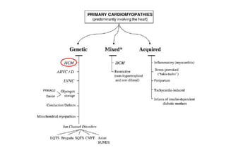
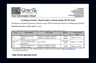
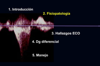
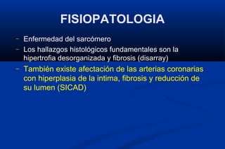
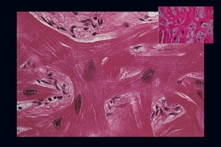
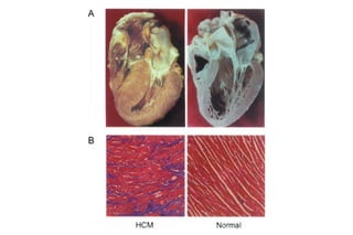
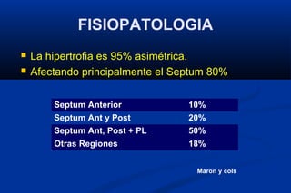
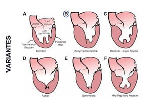
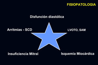
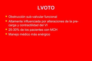
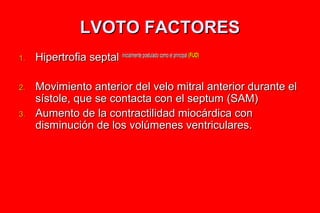
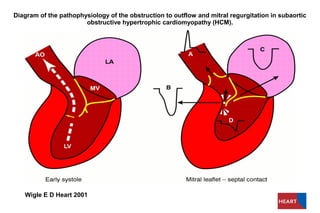
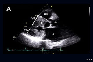
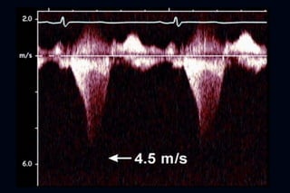
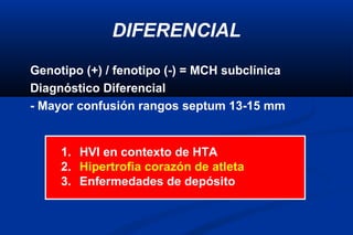
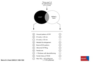
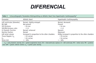
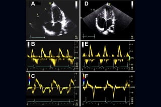
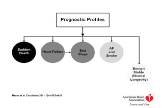
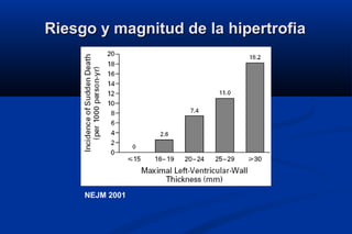
CORE CURRICULUM
MIOCARDIOPATÍA HIPERTRÓFICA
                 Julián Vega Adauy
               Becario Cardiología
1. Introducción
                    2. Fisiopatología


                          3. Hallazgos ECO

              4. Dg diferencial


              5. Manejo
GIOVANNI MARIA LANCISI’S BOOK ON SUDDEN DEATH,
      1707 (Credit: Wellcome Library, London).
─   Teare describió una hipertrofia asimétrica del tabique
    interventricular en 8 pacientes de entre 14 y 44 años de
    edad
    ─   Describió características
         ─   Clínicas
         ─   del EKG
         ─   Anatomopatológicas y la relación con el parentesco
CLASIFICACIÓN


        MIOCARDIOPATÍAS
        MIOCARDIOPATÍAS


PRIMARIAS
PRIMARIAS             SECUNDARIAS
                      SECUNDARIAS
Epidemiología MCH
– Genotipo 0,2% (1/500) en población general
– Cardiopatía hereditaria mas frecuente.
  - Prevalencia 0.1-0.2% en población general

- Mujeres y raza afro-americana aparentemente x 2

- Gran grupo subdiagnósticado
INTRODUCCIÓN
   Se define como una hipertrofia significativa en ausencia de
    causas cardiacas o sistémicas que puedan producirla
    - Se presenta con obstrucción en 1/3 de los casos.
   Tiene un genotipo y fenotipo heterogéneo
   Es resultado de la mutación de proteínas del sarcómero


          Causa mas frecuente de MS en atletas
NOMENCLATURA
─   >80 términos utilizados para describirla
─   IHSS: Idiopathic hypertrophic subaortic stenosis
─   HOCM: hypertrophic obstructive cardiomiopathy
    ─   Elementos confusores pues asumen que la LVOTO es un
        componente invariable
    ─   MCH término acuñado en 1979, incluye ausencia o presencia de
        LVOTO
MUTACION GENETICA
   60-70% de los pacientes con historia familiar de MCH
   30-40% casos esporádicos
   Genotipo (+) / fenotipo (-) = MCH subclinica
   Mutación autosómica dominante en genes que codifican
    proteínas contráctiles del sarcómero
       >1.400 mutaciones en 8 genes
       Myosin heavty chain MYH7 y Myosin binding protein C MYBPC3 = 80%
        mutaciones
       Mutación específica no altera el manejo, pero distinto pronóstico
       Pacientes con >1 mutación (<5%) = enfermedad más severa
A TWO-HIT MODEL TO EXPLAIN VARIABILITY IN HCM PATHOLOGY.


     Wolf C M et al. PNAS 2005
http://www.genedx.com/
1. Introducción
                    2. Fisiopatología


                          3. Hallazgos ECO

              4. Dg diferencial


              5. Manejo
FISIOPATOLOGIA
–   Enfermedad del sarcómero
–   Los hallazgos histológicos fundamentales son la
    hipertrofia desorganizada y fibrosis (disarray)
–   También existe afectación de las arterias coronarias
    con hiperplasia de la intima, fibrosis y reducción de
    su lumen (SICAD)
Small Intramural coronary arteriole dysplasia (SICAD)


  Kwon et al. JACC 2009
FISIOPATOLOGIA
   La hipertrofia es 95% asimétrica.
   Afectando principalmente el Septum 80%


        Septum Anterior           10%
        Septum Ant y Post         20%
        Septum Ant, Post + PL     50%
        Otras Regiones            18%


                                    Maron y cols
VARIANTES
FISIOPATOLOGIA


              Disfunción diastólica


Arritmias - SCD                       LVOTO, SAM


Insuficiencia Mitral             Isquemia Miocárdica
LVOTO
   Obstrucción sub-valvular funcional
   Altamente influenciada por alteraciones de la pre-
    carga y contractilidad del VI
   25-30% de los pacientes con MCH
   Manejo médico más enérgico
LVOTO FACTORES
1.   Hipertrofia septal inicialmente postulado como el principal (FIJO)

2.   Movimiento anterior del velo mitral anterior durante el
     sístole, que se contacta con el septum (SAM)
3.   Aumento de la contractilidad miocárdica con
     disminución de los volúmenes ventriculares.
Diagram of the pathophysiology of the obstruction to outflow and mitral regurgitation in subaortic
                       obstructive hypertrophic cardiomyopathy (HCM).


   Wigle E D Heart 2001
LVOTO


1/3 de pacientes en cada grupo
LVOTO y SV


P. Elliot, EHJ 2006
SAM
   No es un hallazgo patognomónico de la MCH
SAM


JACC 1993
J Am Coll Cardiol Img. 2010;
DISFUNCIÓN DIASTÓLICA

─   Puede presentarse hasta en el 80% de los pacientes
    con miocardiopatía hipertrófica
─   Puede verse en pacientes sin gran HVI, pero que
    tienen el genotipo de MCH
─   Subclínica, TDI hallazgo precos para estratificar
─   No hay clara relación entre la severidad de la
    hipertrofia y la severidad de la disfunción diastólica
DISFUNCIÓN AUTONÓMICA
   Durante el ejercicio 25% tiene respuesta anormal de la presión
    arterial
    -   Caída PAS
    -   Falla en aumentar la PAS > 20 mmHg
    -   Asociado a mal pronóstico
    -   Relacionada a LVOTO y vasodilatación relacionada con ejercicio
ISQUEMIA MIOCÁRDICA
─   Generalmente no relacionada con CAD
    ─   Mismatch entre oferta-demanda
    ─   Aumento de demanda por HVI
─   Compromiso de Q coronario
    ─   (hipertrofia de la media, arterias intramurales)
─   Inducir arritmias y MS
─   Hasta 40% de isquemia detectada por Medicina Nuclear
ISQUEMIA MIOCÁRDICA
   No es parte de la evaluación rutinaria de la MCH en pacientes
    asintomáticos
   Recomendación en pacientes sintomáticos con probabilidad media
    de CAD
   Isquemia de origen micro-vascular
       Asociación con CAD hasta un 20%
       Reportado asociación con CAD severa hasta 26% Sorajja P, Circulation 2003
       MCH con indicación Cx, 25% tienen presencia de CAD que requirió CRM
       15% presencia de puentes musculares en ADA en Coronariografía
       (40% reportada en serie de anatomía patológica)
INSUFICIENCIA MITRAL
–   Factor principal en producir disnea
–   Distorsión del aparato mitral producto del SAM 2rio a LVOTO
    –   Jet de predominio lateral y posterior (97%) Yu, E. H. C. et al. J Am Coll Cardiol 2000
–   Jet de Predominio anterior, descartar alteración 1ria
    –   10-20% IM independiente de SAM
          –   PVM, Ruptura CT, Fibrosis del velo anterior, Calcificación anular, inserción
              anómala de MP
–   Severidad en algunos casos correlación con magnitud de LVOT
The relationship between the LVOT gradient and the mitral regurgitation jet area


                                                        Yu, E. H. C. et al. J Am Coll Cardiol 2000
1. Introducción
                    2. Fisiopatología


                          3. Hallazgos ECO

              4. Dg diferencial


              5. Manejo
Hallazgos ecocardiográficos
   La variante más común (hipertrofia septal asimétrica)
       Hipertrofia del septum en toda su longitud a costa de la
        cavidad
       Razón de pared septal/posterior > 1,3
       Curvatura reversa del septum
   Hipertrofia de predominio “superior” es más frecuente
    en HTA y con envejecimiento
PLAX
Hallazgos ecocardiográficos
   Una característica típica es el impacto del aumento del
    septum en el Doppler del TSVI
   Patrón con contorno de daga
       Dagger shaped spectral Doppler pattern
       Refleja un peak tardío de flujo sistólico a través del TSVI
CW Doppler TSVI
1. Introducción
                    2. Fisiopatología


                          3. Hallazgos ECO

              4. Dg diferencial


              5. Manejo
DIFERENCIAL
Genotipo (+) / fenotipo (-) = MCH subclínica
Diagnóstico Diferencial
- Mayor confusión rangos septum 13-15 mm


     1. HVI en contexto de HTA
     2. Hipertrofia corazón de atleta
     3. Enfermedades de depósito
Distribution of maximum left ventricular (LV) wall thicknesses in athletic population.


       Maron B J Br J Sports Med 2009
ROLE OF TYPE OF SPORT


Maron B J Br J Sports Med 2009
Maron B J Heart 2005;91:1380-1382
DIFERENCIAL
MANEJO
Maron et al. Circulation 2011;124:e783-e831
TRATAMIENTO MEDICO
   No existen estudios RCT, todas las recomendaciones son a
    partir de juicio clínico
    - Terapia apunta a: disminuir LVOTO y SAM
   B-Bloqueadores y agentes inotropos negativos (BCC)
    -   Primera línea: BB si LVOTO, 70% de efectividad en alivio sintomático.
        Evitar agentes con efecto alfa-bloqueador
    -   - Verapamilo si BB no tolera o no responde, hasta 480 mg/d
    -   Precaución en pacientes con gradientes muy elevados
PRONOSTICO
   Históricamente se reportaba mortalidad 3-7% anual
   Actualmente mortalidad 1% anual
       SV >90% a 5ª
       SV >85% a 10ª
       10-15% evoluciona a MCD
Ho C Y Circulation 2012
ESTRATIFICACION DE RIESGO PARA SCD
   Difícil objetivar el riesgo de SCD en prevención 1ria
   Evaluación inicial y luego periódica (anual)
   Trabajo de Maron et al. 2007 sugirió los principales FR
       55% de los pacientes no tienen FR
       10-20% tiene alguno de los FR para SCD
       Factores dinámicos (Gradiente)
       FR con efecto no sumativo, no establecido, sinergismo?
Perry Elliott. EHJ 2006
Estratificación de riesgo de MS, antes 2006

   Criterios Mayores
       Historia de MS recuperada.
       TV sostenida espontanea.
       Historia de MS en un familiar de primer grado.


   Criterios menores
       Hipertrofia mayor a 30 mm.
       TVNS
       Síncope no explicado por otra causa.
       Hipotensión en ejercicio.
       Defectos de perfusión a la RNM.
Riesgo y magnitud de la hipertrofia


     NEJM 2001
PREDICTORES DE SCD (AHA 2011)
FACTORES DE RIESGO PARA SCD y Sobrevida


P. Elliot, EHJ 2006
INDICACIONES DE DAI EN MCH
ICD
   Descargas apropiadas en MCH
   P. Primaria 5% por año
   P. Secundaria 11% por año
   En promedio el ICD descarga tras 9 años post-implante
   Paciente de mediana edad (40ª)


    B. Maron, JAMA 2002
Facultad de Medicina
Pontificia Universidad Católica de Chile


   CORE CURRICULUM
 MIOCARDIOPATÍA HIPERTRÓFICA
                            Julián Vega Adauy, Becario Cardiología


                                                                     ABRIL 2013

Miocardiopatia hipertrófica

  • 1.
    CORE CURRICULUM MIOCARDIOPATÍA HIPERTRÓFICA Julián Vega Adauy Becario Cardiología
  • 2.
    1. Introducción 2. Fisiopatología 3. Hallazgos ECO 4. Dg diferencial 5. Manejo
  • 4.
    GIOVANNI MARIA LANCISI’SBOOK ON SUDDEN DEATH, 1707 (Credit: Wellcome Library, London).
  • 5.
    Teare describió una hipertrofia asimétrica del tabique interventricular en 8 pacientes de entre 14 y 44 años de edad ─ Describió características ─ Clínicas ─ del EKG ─ Anatomopatológicas y la relación con el parentesco
  • 6.
    CLASIFICACIÓN MIOCARDIOPATÍAS MIOCARDIOPATÍAS PRIMARIAS PRIMARIAS SECUNDARIAS SECUNDARIAS
  • 8.
    Epidemiología MCH – Genotipo0,2% (1/500) en población general – Cardiopatía hereditaria mas frecuente. - Prevalencia 0.1-0.2% en población general - Mujeres y raza afro-americana aparentemente x 2 - Gran grupo subdiagnósticado
  • 9.
    INTRODUCCIÓN  Se define como una hipertrofia significativa en ausencia de causas cardiacas o sistémicas que puedan producirla - Se presenta con obstrucción en 1/3 de los casos.  Tiene un genotipo y fenotipo heterogéneo  Es resultado de la mutación de proteínas del sarcómero Causa mas frecuente de MS en atletas
  • 11.
    NOMENCLATURA ─ >80 términos utilizados para describirla ─ IHSS: Idiopathic hypertrophic subaortic stenosis ─ HOCM: hypertrophic obstructive cardiomiopathy ─ Elementos confusores pues asumen que la LVOTO es un componente invariable ─ MCH término acuñado en 1979, incluye ausencia o presencia de LVOTO
  • 12.
    MUTACION GENETICA  60-70% de los pacientes con historia familiar de MCH  30-40% casos esporádicos  Genotipo (+) / fenotipo (-) = MCH subclinica  Mutación autosómica dominante en genes que codifican proteínas contráctiles del sarcómero  >1.400 mutaciones en 8 genes  Myosin heavty chain MYH7 y Myosin binding protein C MYBPC3 = 80% mutaciones  Mutación específica no altera el manejo, pero distinto pronóstico  Pacientes con >1 mutación (<5%) = enfermedad más severa
  • 14.
    A TWO-HIT MODELTO EXPLAIN VARIABILITY IN HCM PATHOLOGY. Wolf C M et al. PNAS 2005
  • 15.
  • 16.
    1. Introducción 2. Fisiopatología 3. Hallazgos ECO 4. Dg diferencial 5. Manejo
  • 17.
    FISIOPATOLOGIA – Enfermedad del sarcómero – Los hallazgos histológicos fundamentales son la hipertrofia desorganizada y fibrosis (disarray) – También existe afectación de las arterias coronarias con hiperplasia de la intima, fibrosis y reducción de su lumen (SICAD)
  • 20.
    Small Intramural coronaryarteriole dysplasia (SICAD) Kwon et al. JACC 2009
  • 22.
    FISIOPATOLOGIA  La hipertrofia es 95% asimétrica.  Afectando principalmente el Septum 80% Septum Anterior 10% Septum Ant y Post 20% Septum Ant, Post + PL 50% Otras Regiones 18% Maron y cols
  • 23.
  • 24.
    FISIOPATOLOGIA Disfunción diastólica Arritmias - SCD LVOTO, SAM Insuficiencia Mitral Isquemia Miocárdica
  • 25.
    LVOTO  Obstrucción sub-valvular funcional  Altamente influenciada por alteraciones de la pre- carga y contractilidad del VI  25-30% de los pacientes con MCH  Manejo médico más enérgico
  • 26.
    LVOTO FACTORES 1. Hipertrofia septal inicialmente postulado como el principal (FIJO) 2. Movimiento anterior del velo mitral anterior durante el sístole, que se contacta con el septum (SAM) 3. Aumento de la contractilidad miocárdica con disminución de los volúmenes ventriculares.
  • 27.
    Diagram of thepathophysiology of the obstruction to outflow and mitral regurgitation in subaortic obstructive hypertrophic cardiomyopathy (HCM). Wigle E D Heart 2001
  • 28.
  • 29.
    LVOTO y SV P.Elliot, EHJ 2006
  • 30.
    SAM  No es un hallazgo patognomónico de la MCH
  • 31.
  • 32.
    J Am CollCardiol Img. 2010;
  • 33.
    DISFUNCIÓN DIASTÓLICA ─ Puede presentarse hasta en el 80% de los pacientes con miocardiopatía hipertrófica ─ Puede verse en pacientes sin gran HVI, pero que tienen el genotipo de MCH ─ Subclínica, TDI hallazgo precos para estratificar ─ No hay clara relación entre la severidad de la hipertrofia y la severidad de la disfunción diastólica
  • 34.
    DISFUNCIÓN AUTONÓMICA  Durante el ejercicio 25% tiene respuesta anormal de la presión arterial - Caída PAS - Falla en aumentar la PAS > 20 mmHg - Asociado a mal pronóstico - Relacionada a LVOTO y vasodilatación relacionada con ejercicio
  • 35.
    ISQUEMIA MIOCÁRDICA ─ Generalmente no relacionada con CAD ─ Mismatch entre oferta-demanda ─ Aumento de demanda por HVI ─ Compromiso de Q coronario ─ (hipertrofia de la media, arterias intramurales) ─ Inducir arritmias y MS ─ Hasta 40% de isquemia detectada por Medicina Nuclear
  • 36.
    ISQUEMIA MIOCÁRDICA  No es parte de la evaluación rutinaria de la MCH en pacientes asintomáticos  Recomendación en pacientes sintomáticos con probabilidad media de CAD  Isquemia de origen micro-vascular  Asociación con CAD hasta un 20%  Reportado asociación con CAD severa hasta 26% Sorajja P, Circulation 2003  MCH con indicación Cx, 25% tienen presencia de CAD que requirió CRM  15% presencia de puentes musculares en ADA en Coronariografía  (40% reportada en serie de anatomía patológica)
  • 37.
    INSUFICIENCIA MITRAL – Factor principal en producir disnea – Distorsión del aparato mitral producto del SAM 2rio a LVOTO – Jet de predominio lateral y posterior (97%) Yu, E. H. C. et al. J Am Coll Cardiol 2000 – Jet de Predominio anterior, descartar alteración 1ria – 10-20% IM independiente de SAM – PVM, Ruptura CT, Fibrosis del velo anterior, Calcificación anular, inserción anómala de MP – Severidad en algunos casos correlación con magnitud de LVOT
  • 38.
    The relationship betweenthe LVOT gradient and the mitral regurgitation jet area Yu, E. H. C. et al. J Am Coll Cardiol 2000
  • 39.
    1. Introducción 2. Fisiopatología 3. Hallazgos ECO 4. Dg diferencial 5. Manejo
  • 40.
    Hallazgos ecocardiográficos  La variante más común (hipertrofia septal asimétrica)  Hipertrofia del septum en toda su longitud a costa de la cavidad  Razón de pared septal/posterior > 1,3  Curvatura reversa del septum  Hipertrofia de predominio “superior” es más frecuente en HTA y con envejecimiento
  • 41.
  • 43.
    Hallazgos ecocardiográficos  Una característica típica es el impacto del aumento del septum en el Doppler del TSVI  Patrón con contorno de daga  Dagger shaped spectral Doppler pattern  Refleja un peak tardío de flujo sistólico a través del TSVI
  • 44.
  • 49.
    1. Introducción 2. Fisiopatología 3. Hallazgos ECO 4. Dg diferencial 5. Manejo
  • 50.
    DIFERENCIAL Genotipo (+) /fenotipo (-) = MCH subclínica Diagnóstico Diferencial - Mayor confusión rangos septum 13-15 mm 1. HVI en contexto de HTA 2. Hipertrofia corazón de atleta 3. Enfermedades de depósito
  • 51.
    Distribution of maximumleft ventricular (LV) wall thicknesses in athletic population. Maron B J Br J Sports Med 2009
  • 52.
    ROLE OF TYPEOF SPORT Maron B J Br J Sports Med 2009
  • 53.
    Maron B JHeart 2005;91:1380-1382
  • 54.
  • 56.
  • 57.
    Maron et al.Circulation 2011;124:e783-e831
  • 58.
    TRATAMIENTO MEDICO  No existen estudios RCT, todas las recomendaciones son a partir de juicio clínico - Terapia apunta a: disminuir LVOTO y SAM  B-Bloqueadores y agentes inotropos negativos (BCC) - Primera línea: BB si LVOTO, 70% de efectividad en alivio sintomático. Evitar agentes con efecto alfa-bloqueador - - Verapamilo si BB no tolera o no responde, hasta 480 mg/d - Precaución en pacientes con gradientes muy elevados
  • 59.
    PRONOSTICO  Históricamente se reportaba mortalidad 3-7% anual  Actualmente mortalidad 1% anual  SV >90% a 5ª  SV >85% a 10ª  10-15% evoluciona a MCD
  • 60.
    Ho C YCirculation 2012
  • 61.
    ESTRATIFICACION DE RIESGOPARA SCD  Difícil objetivar el riesgo de SCD en prevención 1ria  Evaluación inicial y luego periódica (anual)  Trabajo de Maron et al. 2007 sugirió los principales FR  55% de los pacientes no tienen FR  10-20% tiene alguno de los FR para SCD  Factores dinámicos (Gradiente)  FR con efecto no sumativo, no establecido, sinergismo?
  • 62.
  • 63.
    Estratificación de riesgode MS, antes 2006  Criterios Mayores  Historia de MS recuperada.  TV sostenida espontanea.  Historia de MS en un familiar de primer grado.  Criterios menores  Hipertrofia mayor a 30 mm.  TVNS  Síncope no explicado por otra causa.  Hipotensión en ejercicio.  Defectos de perfusión a la RNM.
  • 64.
    Riesgo y magnitudde la hipertrofia NEJM 2001
  • 65.
  • 66.
    FACTORES DE RIESGOPARA SCD y Sobrevida P. Elliot, EHJ 2006
  • 67.
  • 68.
    ICD  Descargas apropiadas en MCH  P. Primaria 5% por año  P. Secundaria 11% por año  En promedio el ICD descarga tras 9 años post-implante  Paciente de mediana edad (40ª) B. Maron, JAMA 2002
  • 69.
    Facultad de Medicina PontificiaUniversidad Católica de Chile CORE CURRICULUM MIOCARDIOPATÍA HIPERTRÓFICA Julián Vega Adauy, Becario Cardiología ABRIL 2013

Notas del editor

  • #5 Giovanni Maria Lancisi’s book on sudden death, 1707 (Credit: Wellcome Library, London). The most noteworthy Renaissance contribution came from Giovanni Maria Lancisi (1654–1720), physician to Pope Clement XI, who was asked to investigate an apparent epidemic of sudden deaths occurring in Rome in 1705. Lancisi performed autopsies and published his treatise on fatal illness, De subitaneis mortibus, where he reported deaths due to hypertrophy, dilatation and valve defects ( fig 1 ). 3 4 His book is important not only because it provides convincing evidence that myocardial hypertrophy was associated with sudden death, but also because it represents a shift away from the classic humoral theory towards clinico-pathological observation. The fact that the Vatican commissioned this work illustrates the growing interest in and acceptance of science by the Church. Over and above his work on sudden death, Lancisi was the first to mention the hereditary tendency to cardiac disease (he describes a family with disease recurrence in four generations) and made one of the first attempts to classify the cardiac diseases then recognised. 4 5 He was well attuned to non-medical issues and there is an obvious humanitarian thread to his work. Interestingly he cautions, “Not every patient who is threatened with danger of sudden death should be warned of this”.4
  • #15 A two-hit model to explain variability in HCM pathology. Incorporation of mutant proteins produces sarcomere defects that trigger abnormal Ca2+ signaling that stimulates myocyte hypertrophy and myocyte disarray and increases cardiac fibrosis (19, 20). The severity of HCM pathology is modified by “ second hits ” or somatic events (potentially exercise, diet, and local hemodynamic factors) that exacerbate hypertrophy, disarray, and fibrosis. An association between even modest increases in LVWT and arrhythmia vulnerability in MHC403/+ mice suggests a shared mechanistic pathway.
  • #20 Pathology of hypertrophic cardiomyopathy A. Post-mortem examination of the heart from an individual ith HCM demonstrating massive asymmetric left ventricular hypertrophy, with associated reduction in left ventricular cavity size, compared to a normal heart. B. Histopathology of heart sections stained with Massons trichrome showing significant myofibre disarray and interstitial fibrosis in HCM.
  • #21 Small Intramural Dysplastic Coronary Arteriole
  • #22 Small intramural coronary artery shows a narrowed lumen due to disordered smooth muscle hyperplasia in the intima and media. There is increased deposition of mucopolysaccharide (blue green) in the intima and collagen (yellow) in the adventitia.
  • #28 Diagram of the pathophysiology of the obstruction to outflow and mitral regurgitation in subaortic obstructive hypertrophic cardiomyopathy (HCM). Early systole (left): The left ventricular outflow tract is narrowed by the ventricular septal hypertrophy and the anterior displacement of the papillary muscles and the mitral leaflets. The point of coaptation of the elongated mitral leaflets occurs in the body of the leaflets, rather than at the tips, as is normal. That part of the anterior leaflet beyond the coaptation point is carried anteriorly and superiorly (systolic anterior motion, arrow) by venturi and/or drag forces and results in mitral leaflet – septal contact, causing the subaortic obstruction (indicated by the converging and diverging lines, right). Mitral leaflet – septal contact (right): The systolic anterior motion of the anterior leaflet results in a failure of coaptation of the mitral leaflets and the onset of mitral regurgitation, which is directed posteriorly into the left atrium, through the funnel shaped interleaflet gap. The length and mobility of the posterior leaflet may also affect the size of this gap, and hence the degree of mitral regurgitation. A, B, C, and D indicate Doppler velocity recordings throughout systole in the ascending aorta (flow toward transducer), at the level of mitral leaflet – septal contact (B), in the left atrium (C), and near the apex of the left ventricle (D). In B, C, and D, flow is away from the transducer. Reduced forward flow in the presence of the obstruction is indicated by the shape of the aortic velocity waveform (A) and the smaller aortic arrow. Peak velocities recorded at B correlate accurately with the simultaneously measured pressure gradient, whereas late peaking velocities at D do not. AO, aorta; LA, left atrium, LV, left ventricle, MV, mitral valve. Adapted from Wigle et al.7
  • #33 En-face Views of Aortic Valve Showing Unobstructed LVOT Without (A) and With (B) SAM; LVOT Obstruction Due to SAM is Seen Through Aortic Valve Opening (B) A (en-face, short-axis view) shows a normal left ventricular outflow tract (LVOT) visible during systole through the opening of the aortic valve with systolic blood pressure &gt;120 mm Hg. On the right (B) is the same patient with systolic blood pressure &lt;100 mm Hg where the aortic valve opening is visible, but the left ventricular outflow tract is completely obstructed with the anterior mitral leaflet touching the interventricular septum. A clear visualization of a normally moving anterior mitral leaflet is seen in Online Video 1 and systolic anterior motion (SAM) is visible in Online Video 2 with the anterior mitral leaflet coming in contact with the interventricular septum.
  • #42 Figure 1 Characteristic Echocardiographic Features of Obstructive HCM (A) Parasternal long-axis view depicting severe asymmetric septal hypertrophy and systolic anterior mitral valve motion (arrowhead) ; (B) M-mode across the mitral leaflet...
  • #43 Figure 1 Characteristic Echocardiographic Features of Obstructive HCM (A) Parasternal long-axis view depicting severe asymmetric septal hypertrophy and systolic anterior mitral valve motion (arrowhead) ; (B) M-mode across the mitral leaflet...
  • #45 Figure 2. Contrasting the CW Doppler Through the Orifice of Patients With Aortic Stenosis and Obstructive HCMIn aortic stenosis the orifice remains relatively fixed in systole. In hypertrophic cardiomyopathy (HCM) obstruction begets more obstruction, and the orifice narrows through systole. The left ventricular ejection is unobstructed in early systole, and the curve is convex-to-the-left. At the inflection point (long arrow) , mitral-septal contact and obstruction begin. The orifice narrows because of the rising pressure difference; the pressure difference rises because of the decreasing orifice size—an amplifying feedback loop. Hence, the contour shows an increasing acceleration pattern: it is concave-to-the-left (arrowheads) . CW = continuous wave. Adapted, with permission, from Sherrid et al. The amplifying nature of outflow obstruction has significance for the therapy of symptomatic gradients. The longer in systole that the gradient acts, and the longer the duration of mitral-septal contact, the higher the gradient. Successful therapy abolishes or delays SAM (6). The amplifying nature of outflow obstruction also may explain the dramatic changes in gradient that occur with loading conditions, and may explain the sometimes explosive emergence of severe outflow obstruction in the elderly.
  • #46 Figure 7 Typical continuous wave Doppler recording from a patient with dynamic LVOT obstruction in the setting of hypertrophic obstructive cardiomyopathy. Note the dagger-shaped profile. (Reproduced with permission from Oh et al. &lt;ce:cross-ref re...
  • #47 Figure 1 CW Doppler Echocardiographic Tracing Through the Obstructing Orifice in the LVOT of a Patient With a Gradient of 64 mm Hg The inflection point is shown with a long arrow and the concave-to-the-left contour is shown with arrowheads . ...
  • #48   Demonstration of sudden marked reduction of forward aortic flow. Aortic stenosis velocity profile demonstrating sudden decrease in flow at 230 ms after onset of systole, followed by low-velocity flow (1.2 m/sec) to aortic valve closure at 380 ms. Late peaking velocity profile typical of dynamic obstruction accelerating at 230 ms after onset of systole and continuing through to aortic valve closure. SAM, Systolic anterior motion
  • #49   Demonstration of sudden marked reduction of forward aortic flow. Aortic stenosis velocity profile demonstrating sudden decrease in flow at 230 ms after onset of systole, followed by low-velocity flow (1.2 m/sec) to aortic valve closure at 380 ms. Late peaking velocity profile typical of dynamic obstruction accelerating at 230 ms after onset of systole and continuing through to aortic valve closure. SAM, Systolic anterior motion
  • #52 Distribution of maximum left ventricular (LV) wall thicknesses in a large young male and female athletic population. Two per cent of male and 0% of female athletes have a wall thickness ⩾13 mm in the ambiguous “grey-zone” of athlete’s heart versus hypertrophic cardiomyopathy. Reproduced from Pelliccia et al,25 with permission of the Massachusetts Medical Society.
  • #53 Role of type of sport. Effect of specific sports training on maximum end-diastolic left ventricular (LV) wall thickness (and transverse cavity dimension) in elite athletes, representing 27 different sporting disciplines. L.D., long-distance running; X-country, cross-country.
  • #54   Criteria used to distinguish hypertrophic cardiomyopathy (HCM) from athlete’s heart when the left ventricular (LV) wall thickness is within the shaded “grey zone” of overlap, consistent with both diagnoses. ↓ indicates decreased; LA, left atrial; LVH, left ventricular hypertrophy. Reproduced from Maron et al,11 with permission of American Heart Association.
  • #58 Prognosis profiles for HCM and targets for therapy. AF indicates atrial fibrillation. Modified with permission from Maron et al.10
  • #61 Key components in the clinical management of hypertrophic cardiomyopathy (HCM). CaChBl indicates calcium channel blocker; SCD, sudden cardiac death; HR, heart rate; ICD, implantable cardioverter-defibrillator; HF, heart failure; and CV, cardiovascular.
  • #67 SV según FR (5) P. Elliot, EHJ 2006
  • #68 MODIFICADORES LVOTO &gt; 30 mmHg LGE en CMRI Aneurisma Apical Mutaciones de alto riesgo