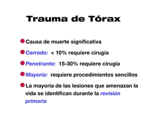
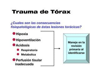
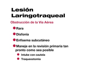
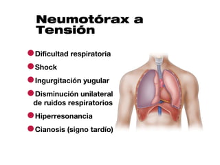
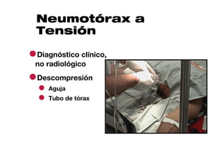
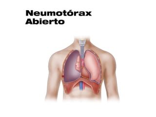
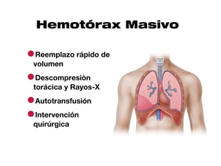
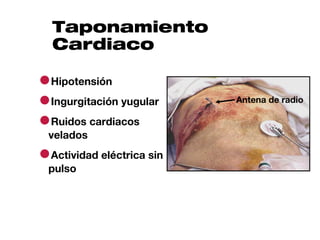
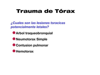
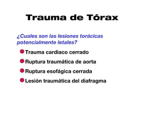
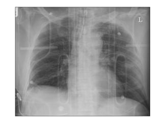
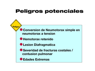
Este documento describe la evaluación y manejo inicial del trauma torácico. Resalta que las lesiones que amenazan la vida como la obstrucción de vía aérea, neumotórax a tensión y hemorragia masiva deben identificarse y tratarse durante la revisión primaria para prevenir complicaciones como hipoxia e hipoperfusión tisular. También destaca que la mayoría de lesiones potencialmente letales como neumotórax simple, contusión pulmonar y hemotorax pueden identificarse con exámen físico, rayos
1. Trauma de
Tórax
Initial Assessment and
Management
2. Caso clínico
● Hombre de 27 años es llevado al
centro de trauma
● Conductor sin cinturón de seguridad,
en impacto frontal a alta velocidad
● Tiempo prolongado de extricación en
la escena del accidente
3. Caso clínico
● Presión arterial: 90/70; frecuencia
cardiaca: 110; frecuencia respiratoria: 36
● Evaluación inicial: Glasgow 15, vía aérea
permeable
¿Que características sugieren que este
paciente puede tener lesiones torácicas?
4. Objetivos
● Identifique y maneje las lesiones
que amenazan la vida encontradas
en la revisión primaria.
● Identifique y maneje las lesiones
potencialmente letales encontradas
durante la revisión secundaria.
6. Traquea y bronquios
Espacios pleurales y parénquima
Mediastino
Diafragma
Torax óseo
Tejidos blandos
Sondas y líneas
7. Trauma de Tórax
●Causa de muerte significativa
●Cerrado: < 10% requiere cirugía
●Penetrante: 15-30% requiere cirugía
●Mayoría: requiere procedimientos sencillos
●La mayoría de las lesiones que amenazan la
vida se identifican durante la revisión
primaria
8. Trauma de Tórax
¿Cuales son las lesiones torácicas que
amenazan la vida de manera inmediata?
●la vía aérea
Lesón laringotraqueal / Obstrucción de
●Neumotorax a tensión
●Neumotorax abierto
●Tórax inestable y contusión pulmonar
●Hemotorax masivo
●Taponamiento cardiaco
9. Trauma de Tórax
¿Cuales son las consecuencias
fisiopatológicas de éstas lesiones torácicas?
●Hipoxia
●Hipoventilación
●Acidosis
Maneje en la
revisión
● Respiratoria primaria al
● Metabolica identificarse
●Perfusión tisular
inadecuada
10. Lesión
Laringotraqueal
Obstrucción de la Vía Aérea
●Rara
●Disfonía
●Enfisema subcutáneo
●Maneje en la revisión primaria tan
pronto como sea posible
● Intube con cautela
● Traqueostomìa
11. Neumotórax a
Tensión
●Dificultad respiratoria
●Shock
●Ingurgitación yugular
●Disminución unilateral
de ruidos respiratorios
●Hiperresonancia
●Cianosis (signo tardío)
12. Neumotórax a
Tensión
●Diagnóstico clínico,
no radiológico
●Descompresión
● Aguja
● Tubo de tórax
16. Tórax Inestable y
Contusión Pulmonar
●Oxígeno
●Reexpandir el
pulmón
●Intubación según
necesario
●Líquidos con buen
juicio
●Analgesia
17. Hemotórax Masivo
●Ruptura de vasos
sistémicos / pulmonares
●Pérdida sanguínea >
1500 mL
●Yugulares ingurgitadas
vs colapsadas
●Shock sin ruidos
respiratorios y/o
matidez a la percusión
20. Taponamiento
Cardiaco
A Asegure la vía aérea
B Ventile y oxigene
C Reanime con volumen
FAST, cirugía
21. Toracotomía de
Reanimación
¿Cuando debo considerar una
toracotomía de reanimación?
22. Toracotomía de
Reanimación
¿Cuando debo considerar una
toracotomía de reanimación?
●Pacientes con lesión torácica
penetrante con AESP al ingreso pueden
ser candidatos
●Cuando hay un cirujano con las
destrezas necesarias
●La toracotomía en urgencias no está
indicada en trauma cerrado con AESP
23. Trauma de Tórax
¿Cuales son las lesiones torácicas
potencialmente letales?
¿Como las identifico?
¿Cuando y como corrijo el problema?
24. Trauma de Tórax
¿Cuales son las lesiones toracicas
potencialmente letales?
●Arbol traqueobronquial
●Neumotorax Simple
●Contusion pulmonar
●Hemotorax
25. Trauma de Tórax
¿Cuales son las lesiones torácicas
potencialmente letales?
●Trauma cardiaco cerrado
●Ruptura traumática de aorta
●Ruptura esofágica cerrada
●Lesión traumática del diafragma
26. Trauma de Tórax
¿Como identifico las lesiones torácicas
potencialmente letales?
●Examen fisico
●Rx de Torax
●Oximetria de pulso
●Gases arteriales
●ECG
31. Trauma Cardiaco
Cerrado
●Gama de lesiones
●ECG anormal / cambios en el
monitor
●Ecocardiograma
●Maneje
● Arritmias
● Perfusión
● Complicaciones
32. Ruptura Traumática
de Aorta
●Mecanismo de
aceleración /
desaceleración
rápida
●Signos
radiológicos
●Alto indice de
sospecha
●Valoracion por
cirugia
36. Ruptura Traumática
de Esófago
●Trauma cerrado vs penetrante
●Golpe epigastrico severo
●Dolor / shock desproporcionados
para la lesión
●Neumotorax o hemotorax
izquierdos sin fractura costal
37. Trauma Esofágico
●Tubo de Torax:
material particulado
●Aire Mediastinal
●Trago de contraste,
esofagoscopia
●Cirugia
38. Trauma
Diafragmatico
●La mayoría diagnosticado
a la izquierda
●Cerrado: desgarros
grandes
●Penetrante:
perforaciones pequeñas
●Rx malinterpretada
●Rx con contraste
●Cirugia
41. Fracturas y Lesiones
Asociadas
Esternon, escapula y costillas
Costillas 1-3
●Fuerza severa
●Lesiones asociadas tienen alto riesgo de muerte
Costillas 4-9
●Contusion pulmonar y neumotorax
Costillas 10-12
●Sospeche lesion abdominal
42. Peligros potenciales
●neumotorax detension
Peligro
Conversion Neumotorax simple en
a
●Hemotorax retenido
●Lesion Diafragmatica
●contusion pulmonar costales /
Severidad de fracturas
●Edades Extremas
43.
44. Resumen
●Comun en pacientes con trauma multiple
●Lesiones que amenazan la vida
●Lesiones potencialmente letales
●Estabilizacion inicial con tecnicas
sencillas en la mayoria de casos
Objetivo: Restaurar intercambio gaseoso y
perfusion normales
Notas del editor
4-1 Title slide Introduce the topic and explain to the students that, based on their preparation for the course, a series of questions will be asked and scenarios are presented. Their participation and responses are expected. This lecture focuses on the ABCD approach to assessment and management of life-threatening and potentially life-threatening chest injuries. Note: This presentation contains case scenarios, didactic information, and key questions. You need to engage the students in discussion, especially when the scenarios are presented. Remember, time management is essential to ensure that all salient points are addressed adequately and to avoid having to hurry through the lecture near the end. X-ray courtesy of Dr. Ray McGlone, Royal Lancaste
4-2 Case Scenario Describe the scenario.
4-3 Case Scenario Describe the scenario. E licit from the students the following answers to the question on the slide: mechanism of injury, significant tachypnea, and moderate tachycardia.
4-4 Objectives Review the objectives as provided on the slide. Emphasize the clinical and other important aspects of the approach to the injured patient with thoracic injuries. Note: This lecture focuses on the ABCDE approach to the initial assessment and management processes of identifying and treating patients who sustain thoracic trauma. For example, the first case scenario presented in this session focuses on the life-threatening airway and breathing injuries of tension pneumothorax, open pneumothorax, and flail chest with pulmonary contusion. Through the discussions you lead, the students will apply knowledge of the principles of initial assessment to achieve a presumptive diagnosis and the ATLS method of treatment.
4-5 Thoracic Trauma Review this information with the students. Emphasize that chest injuries remain a significant cause of morbidity and mortality among trauma patients. Life-threatening injuries associated with thoracic injuries are identified in the primary survey by carefully assessing the patient’s ABCs. Identified injuries usually require simple interventions to secure the airway, reexpand the lung, drain the pleural space, and improve breathing mechanics. Most penetrating wounds to the chest require a thoracostomy tube. A minority of patients with chest injuries require urgent surgical exploration due to bleeding. Use the final point to elicit the six life threatening injuries on the subsequent slide.
4-6 Thoracic Trauma: What are the immediately life-threatening chest injuries? Direct the discussion to prioritize treatment based on the ABCDE algorithm. Emphasize that these life-threatening injuries are treated as they are identified. This list may be used as a tool to briefly contrast the pathophysiology and clinical signs that the student might see. For example, contrast the pathophysiology of hypoxia from airway obstruction with tension pneumothorax. Emphasize that these life-threatening injuries and related problems are resolved as they are discovered. At this point in the lecture, it is not necessary to spend a lot of time on specifics because each injury is discussed in detail during the lecture. Emphasize key recognition factors for each injury listed on this slide.
4-7 Thoracic Trauma: What are the pathophysiologic consequences of these chest injuries? Emphasize the causes of hypoxia and hypoventilation that produce respiratory acidosis. Relate this topic back to the Shock lecture by emphasizing that acidosis is associated with hypovolemia and shock. Stress that inadequate tissue perfusion and ventilation are correctable causes.
4-8 Laryngeotracheal Injury: Airway Obstruction You may use prompts, such as speaking in a hoarse or quiet voice, noisy breathing, and stridor. The students should determine that treatment is to establish a definitive airway. Briefly query the students about the use of endotracheal intubation versus tracheostomy versus surgical cricothyroidotomy. Explain that these skills are discussed and practiced during the skills stations later on the first day of the course.
4-9 Tension Pneumothorax How do I identify a tension pneumothorax? How do I differentiate a tension pneumothorax from cardiac tamponade and hemorrhagic shock? During the discussion of differentiating a tension pneumothorax from cardiac tamponade and hemorrhagic shock, relate that blunt injury is a more likely cause of tension pneumothorax. Cardiac tamponade occurs more often with penetrating trauma. If hypovolemia exists, the patient’s neck veins will not be distended.
4-10 Tension Pneumothorax How do I treat tension pneumothorax? The students should relate that the tension pneumothorax must be immediately decompressed by inserting a needle, followed by insertion of a chest tube. Students should understand that tension pneumothorax is a clinical diagnosis, not confirmed by x-ray. Avoid going into too much detail about the procedures for needle and tube decompression of the chest, but explain that the students have the opportunity to perform these procedures later in the day. Photograph courtesy of Francisco De Salles Collet E. Silva, MD, FACS, Brazil.
4-11 Open Pneumothorax Describe the pathophysiology of an open pneumothorax using the illustration on the slide to explain ineffective ventilation and the rationale and technique for partially occlusive dressing. Use the second slide in the series to emphasize definitive management.
4-12 Open Pneumothorax Photograph courtesy of Francisco De Salles Collet E. Silva, MD, FACS, Brazil
4-13 Flail Chest and Pulmonary Contusion How do I recognize if this patient has a flail chest and pulmonary contusion? This is the only slide addressing the clinical sequelae of flail chest and pulmonary contusion. Emphasize that the problem with the flail chest is related to the patient’s pain and is similar to rib fractures, although it is more severe because of the number of fractures. Further explain that the flail segment serves as a marker for pulmonary contusion, which is the real cause of the patient’s hypoxemia. Photograph used with permission from Trauma.org; Fernando Rodriguez, chest 0021a; Mexico; http://www.trauma.org/imagebank/imagebank.html.
4-14 Flail Chest and Pulmonary Contusion How do I treat the patient with a flail chest and/or pulmonary contusion? Emphasize that the treatment goal is to reexpand the lung, eg, with CPAP (positive pressure) or physiotherapy, and to avoid progressive atelectasis. Analgesia is an important adjunct, but oversedation will promote hypoventilation and atelectasis.
4-15 Massive Hemothorax What is the cause and how do I identify if the patient has a massive hemothorax? Note that this type of injury results in a ‘B’ and ‘C’ problem. X-ray courtesy of Ray McGlone, Royal Lancaster Infirmary; UK
4-16 Massive Hemothorax How do I manage the patient with a massive hemothorax? During the discussion of treatment for a massive hemothorax, emphasize that resuscitation continues in accordance with the shock guidelines. Placement of a thoracostomy tube is essential to expand the lung, and the use of an autotransfusion device is helpful. Medical personnel need to be familiar with the use of the autotransfusion device before it is needed. Most of these devices do not require anticoagulation when the blood is collected. When blood loss continues despite thoracostomy tube drainage and resuscitative efforts are employed, operative intervention is needed.
4-17 Cardiac Tamponade This photograph shows a patient with a parasternal penetrating injury, self-inflicted with a car radio antenna. (Note: The patient’s head is at the bottom of the photograph where one can see the oxygen mask.) Ensure that the students understand that blunt trauma to the chest can cause cardiac tamponade, but that most survivors of cardiac tamponade have an anterior or posterior penetrating wound to the chest. During the discussion about signs and symptoms of the patient with cardiac tamponade, emphasize the fact that not all findings must be present. Explain that cardiac tamponade is suspected if the patient has a penetrating parasternal wound. Clinical signs may include hypotension and dyspnea, or the patient verbalizing that he or she senses he or she is dying. Photograph used with permission of Trauma.org; Frederick Foss; chest0016a; http://www.trauma.org/imagebank/imagebank.html
4-18 Cardiac Tamponade How do I manage the patient with cardiac tamponade? Emphasize the need to remember the ABCs when evaluating and managing a patient with cardiac tamponade. This injury is life-threatening and requires immediate treatment. FAST (focused assessment sonography in trauma) is a useful diagnostic adjunct to use, if available.
4-19 Resuscitative Thoracotomy: When should I consider resuscitative thoracotomy? After asking this question, elicit responses from the students. The students should relate that resuscitative thoracotomy is rarely indicated. Answers appear on the next slide.
4-20 Resuscitative Thoracotomy: When should I consider resuscitative thoracotomy? Patients with a penetrating injury and PEA, especially if the loss of signs of life is recent, are appropriate candidates for resuscitative thoracotomy. A qualified surgeon must be present at the time of the patient’s arrival to determine the need and potential for success of an emergency department resuscitative thoracotomy. Once the chest is opened, bleeding must be controlled.
4-21 Thoracic Trauma Key Questions Review these key questions with the students, explaining that these are the second set of questions for consideration as the patient is being reassessed (ABCDEs). These questions are useful for discussion during the presentation pertaining to the secondary survey. Encourage the students to produce a list of potentially life-threatening chest injuries.
4-22 Thoracic Trauma: What are the potentially life-threatening chest injuries? After the students identify the potentially life-threatening chest injuries, ask how these injuries can threaten life and/or the pathophysiology associated with each injury. Emphasize that simple pneumothorax and pulmonary contusion are the most common thoracic injuries. Subject is continued on the next slide.
4-23 Thoracic Trauma: What are the potentially life-threatening chest injuries? After the students identify the potentially life-threatening chest injuries, ask how these injuries can threaten life and/or the pathophysiology associated with each injury. Emphasize that simple pneumothorax and pulmonary contusion are the most common thoracic injuries.
4-24 Thoracic Trauma: How do I identify potentially life-threatening thoracic injuries? Remember, this list is not exhaustive but reflects the primary survey.
4-25 Tracheobronchial Tree Injury How do I identify and treat a tracheobronchial injury? Explain that a strong suspicion of a tracheobronchial injury should be raised if the lung does not properly inflate after insertion of a chest tube, or if there is a persistent air leak after chest thoracostomy. The students should indicate that a bronchoscopy may be needed after the patient is completely assessed, and that a tracheobronchial injury requires operative repair.
4-26 Simple Pneumothorax How do I identify and treat a simple pneumothorax? The students should understand that a pneumothorax can occur after blunt and penetrating injury to the chest, although it may not be apparent on physical examination. After describing how to identify and treat this injury, ask about the usefulness of obtaining a chest x-ray, which can help identify this injury. Remind students of the need to assess for subcutaneous emphysema. The presence of decreased breath sounds is not always a helpful indicator for a pneumothorax, especially if the patient is hyperventilating from pain or is in shock. The students should know that this injury is treated with tube thoracostomy.
4-27 Pulmonary Contusion How do I identify and treat a pulmonary contusion? A pulmonary contusion can be mild to severe and may cause very little hypoxia to severe hypoxia. The diagnosis can be confirmed by a chest x-ray or a CT scan of the chest. Most pulmonary contusions increase in size and severity after fluid resuscitation. The students should know that treatment includes normovolemia and maneuvers to maintain lung volumes. X-ray courtesy of Ray McGlone, Royal Lancaster Infirmary, UK
4-28 Hemothorax How do I identify and treat a hemothorax? The students should relate that a hemothorax is suspected in cases of hypotension without obvious sources of blood loss or when there are decreased breath sounds and dullness to percussion over a hemithorax. Emphasize that hemothorax is a common source of shock. Hemothorax is confirmed primarily by chest x-ray. The students should relate that initial treatment is tube thoracostomy. Autotransfusion also may be useful. X-ray courtesy of Ray McGlone, Royal Lancaster Infirmary, UK
4-29 Blunt Cardiac Injury How do I identify and treat a patient with blunt cardiac injury? The students should relate that blunt cardiac injury is rare. The patient with blunt cardiac injury usually presents with an abnormal ECG within the first 24 hours from injury. Ask the students about mechanism of injury. They should respond that it results from a direct blow to the chest and heart that affects the cardiac conduction system. Pitfalls include the elderly patient with myocardial ischemia who may already have an abnormal cardiac rhythm. Caveat: dysrhythmias that do not compromise blood pressure may not require treatment.
4-30 Traumatic Aortic Disruption How do I identify and treat traumatic aortic disruption? The students should know that a patient involved in a rapid deceleration or acceleration situation is at risk for an aortic injury. The students also should relate a high index of suspicion for an aortic disruption when the x-ray shows a widened mediastinum. Surgical consultation is necessary. Adjuncts to confirm the diagnosis are on the next slide. X-ray used with permission of Trauma.org; Dr. Hairul Ahmad, widened mediastinum vasc0003a, http://www.trauma.org/imagebank/imagebank.html .
4-31 Traumatic Aortic Disruption How do I confirm the diagnosis of an aortic disruption? Aortogram used with permission of Trauma.org, Dr. Hairul Ahmad, vasc0003b; and helical CT used with permission of Trauma.org, Dr. Luis Filipe Pinheiro, vasc0007b.
4-32 Blunt Esophageal Rupture How do I identify an esophageal injury? Explain that esophageal injury is rare from blunt trauma and is more common from a penetrating injury, especially if the injuring agent is a knife. The students should describe the mechanism for blunt esophageal injury as a severe blow to the upper abdomen that causes a forceful expulsion of gastric contents into the esophagus. An esophageal injury should be suspected in any patient who has received a severe blow to the epigastrium and is in shock or pain out of proportion to the apparent injury. Esophageal injury also should be suspected if the patient has a left pneumothorax or hemothorax without a related rib fracture.
4-33 Esophageal Injury How do I identify and treat an esophageal injury? Students should identify the clinical signs of esophageal injury, including particulate matter in the chest tube and mediastinal air seen on a lateral chest x-ray. Esophageal injury can be diagnosed by contrast swallow or esophagoscopy. Wide drainage of the pleural space and mediastinum with direct operative repair of the injury is the method of treatment. The x-ray shows esophageal perforation with mediastinal air. X-ray used with permission of Trauma.org, Dr. Harry Voesten, chest0027, http:/www.trauma.org/imagebank/imagebank.html
4-34 Diaphragmatic Injury How do I identify and treat diaphragmatic rupture? Ask the students about causes of a diaphragmatic rupture. They should relate that it takes a significant transmission of force to rupture the diaphragm. Explain that diaphragmatic rupture occurs with greater frequency on the left. An elevated diaphragm on the chest x-ray should raise the suspicion for a rupture diaphragm. Explain that an elevated hemidiaphragm can be misinterpreted as a hemothorax. Diaphragmatic rupture requires an operation to repair the injury. X-ray courtesy of Ray McGlone, Royal Lancaster Infirmary, UK.
4-35 Traumatic Asphyxia What is traumatic asphyxia and how do I manage it? Explain that traumatic asphyxia is commonly associated with the acute, temporary compression of the superior vena cava. Venous return is impaired from the upper torso and head. Petechiae occur, as a well brain edema. Most cases are mild, with the patient recovering after the weight is removed from the chest and the head is elevated. Associated injuries must be treated. You may use the photograph to summarize the various signs and symptoms of this injury. Note the area of plethora that begins just above the nipple line and especially of the face. The patient’s eyes are bloodshot. Photograph courtesy of Kimball I. Maull, MD, FACS, USA.
4-36 Subcutaneous Emphysema What type of injury should I suspect with this patient, and how do I treat it? The students should suspect subcutaneous emphysema in this patient, which can be a disturbing visual sign in some patients. This appearance usually indicates an airway injury that may or may not require operative intervention. A blast injury might be another cause. The first step in confirming the diagnosis is to evaluate a chest x-ray or a CT of the chest and identify a pneumothorax. If present, insertion of a chest tube is needed to reexpand the lung. Caution the students about applying an occlusive dressing around the chest tube in this type of patient to avoid the risk of increasing the patient’s subcutaneous emphysema. If the lung can be expanded, this finding is self-limited.
4-37 Fractures and Associated Injuries What associated injuries should I suspect and assess for when my patient has fractures of the chest wall? Explain that the location of the fracture provides clues about other possible injuries. The students should understand that it requires a significant force to fracture ribs 1-3. Patients with these fractures commonly have many associated injuries and are at a higher risk for mortality. Fractures of ribs 4-9 are commonly associated with flail chest, pulmonary contusion, and pneumothorax. Lower rib fractures are associated with intraabdominal injury. Time permitting, review diaphragmatic excursion and its relationship to intraabdominal injuries and lower thoracic injuries.
4-38 Pitfalls What pitfalls should I avoid? Review the pitfalls as provided on the slide. Application of an occlusive dressing over a wound associated with a simple pneumothorax can produce a tension pneumothorax. Attention to chest tube placement and thorough evacuation of the pleural space is important to reduce the risk of a retained hemothorax and infectious complication. Adequate pain relief must be achieved to avoid complications from rib fractures. The elderly with comorbidities are at high risk for complications if adequate ventilation and lung expansion are not maintained.
4-39 Questions? Ask for questions from the students and then pause, allowing the students adequate time to form and ask their questions.
4-40 Summary Use these bulleted items to summarize the presentation. Chest injuries are common in the multiply injured patient. The ABCDE approach is used to identify life-threatening and potentially life-threatening injuries. Initial stabilization requires simple maneuvers, eg, endotracheal intubation and tube thoracostomy. The goal of treating patients with chest injuries is to establish normal gas exchange and normal hemodynamics.